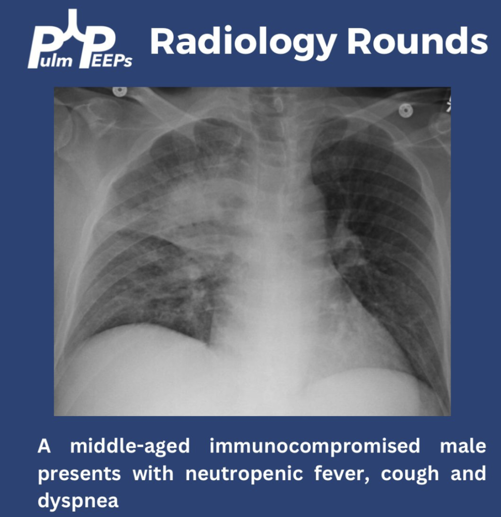
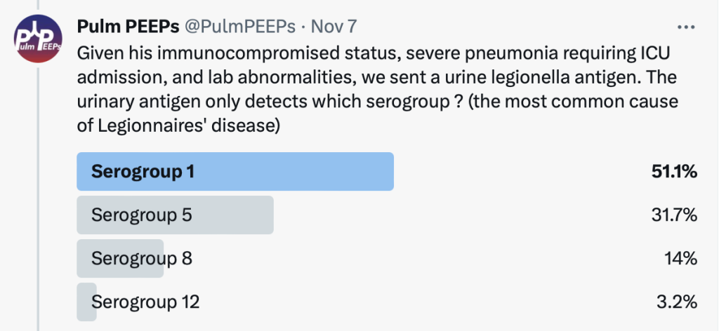
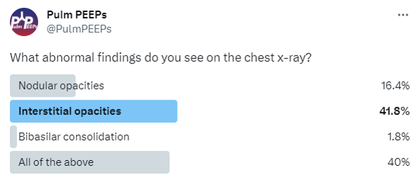
We have a middle-aged immunocompromised male presenting with neutropenic fevers, progressive cough and dyspnea. He has no sick contacts or recent travel.
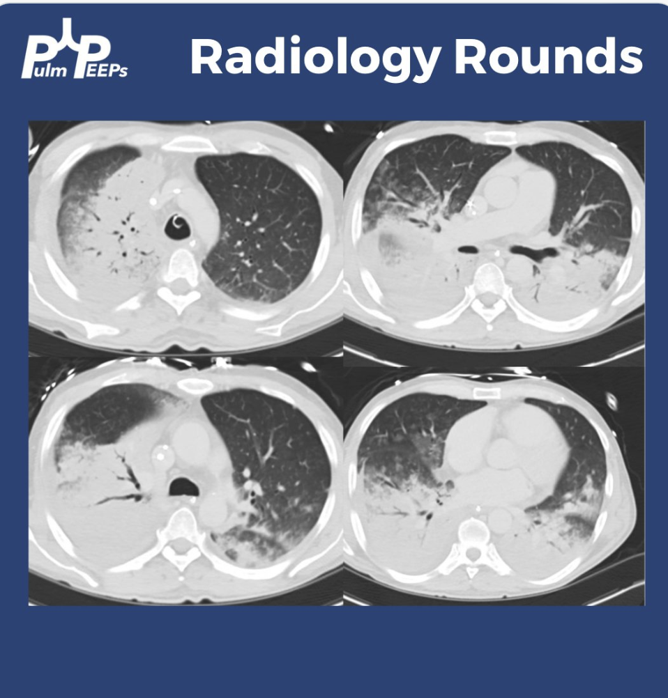
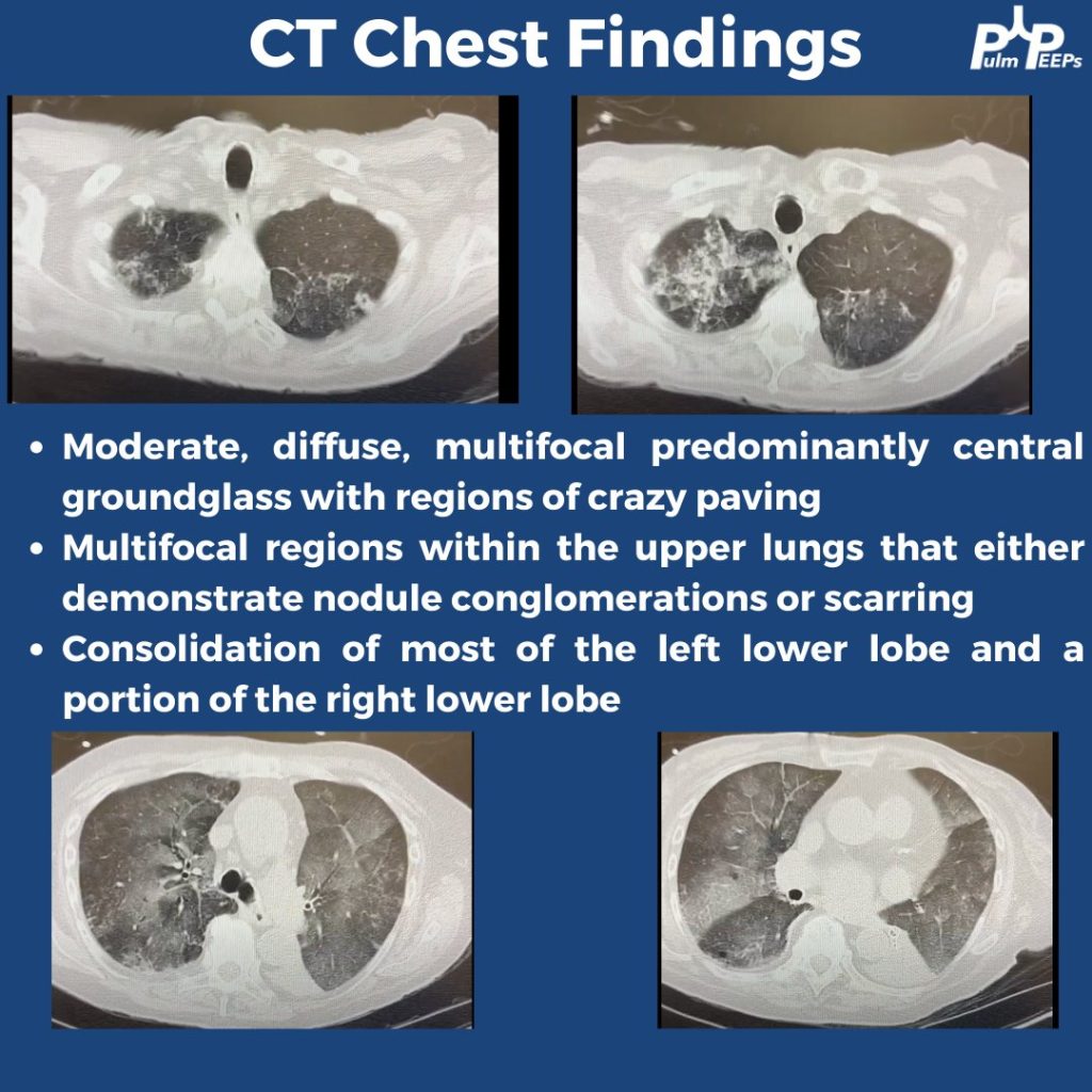
He was found to have primarily right upper alveolar opacities and blunting of the right costophrenic angle. He rapidly decompensated with acute hypoxemic respiratory failure requiring mechanical ventilation. A CT chest showed dense consolidations with air bronchograms.
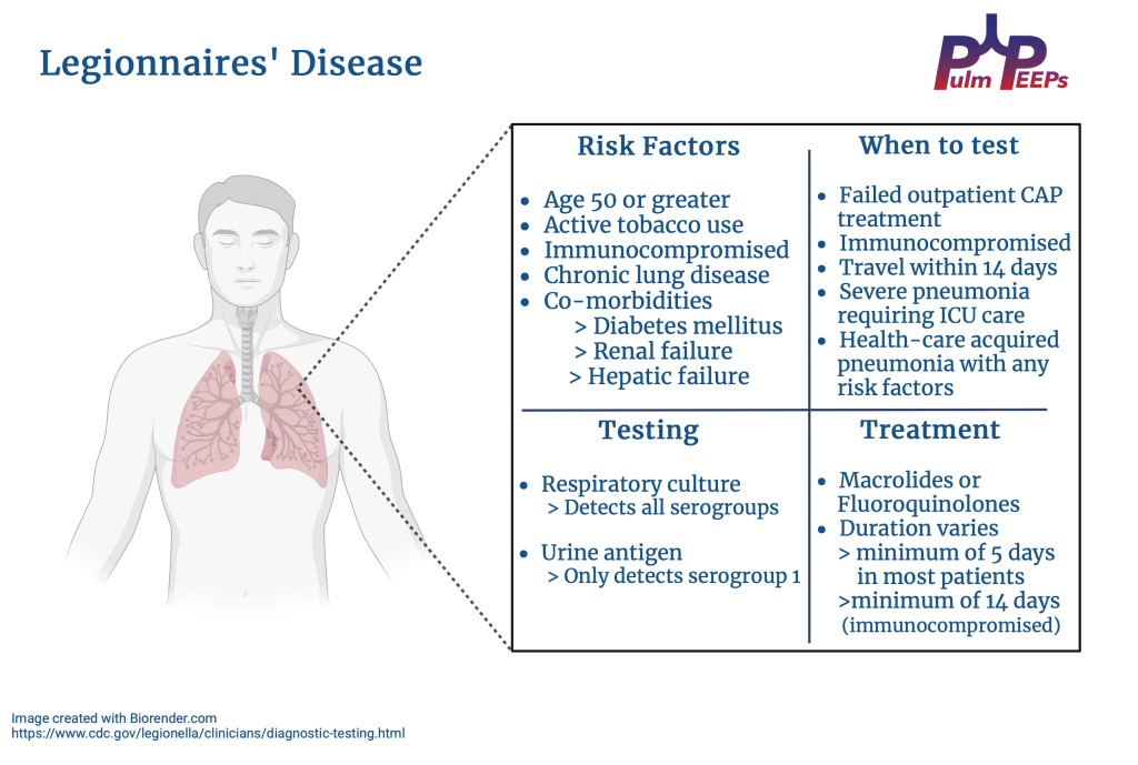
The urinary antigen and sputum culture were positive for Legionella and the patient was continued on Macrolide therapy. See our infographic for high-yield teaching points for Legionnaires’ Disease
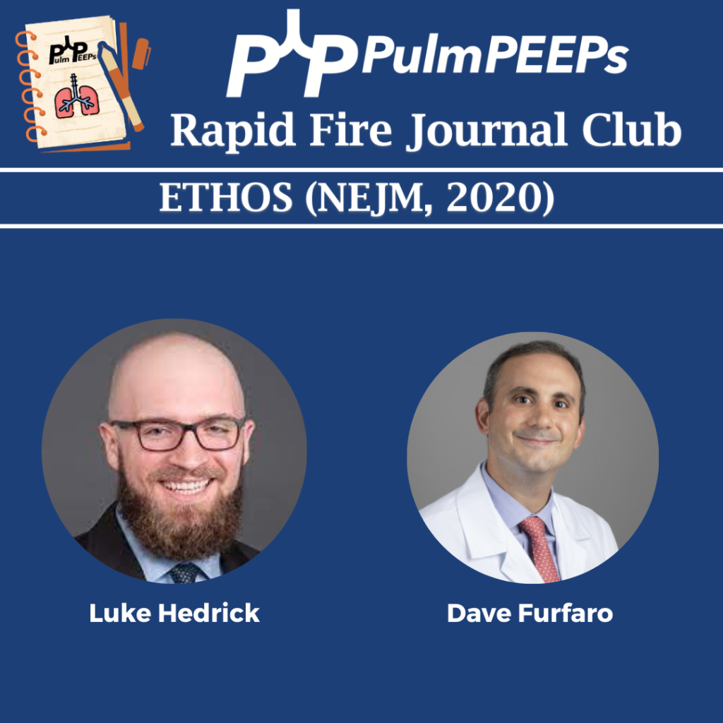
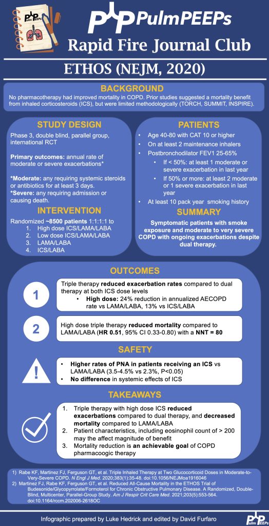
Rapid Fire Journal Club returns with a deep dive into the 2020 ETHOS Trial published in The New England Journal of Medicine examining triple therapy for moderate to severe COPD. Pulm PEEPs Associate Editor Luke Hedrick takes us through this fascinating study and breaks down some of the intricacies.
Article and Reference
Today we’re talking about the 2020 ETHOS Trial in NEJM
This week on Pulm PEEPs, we are excited to be cross-posting an episode that Dave Furfaro did on the ATS Breathe Easy Podcast. Listen to hear a discussion about the best way to create a positive learning environment in the ICU, and how to effectively prepare bedside teaching for learners of all levels.
Meet The Host
Matthew Stutz hosted this episode of the ATS Breathe Easy Podcast. He is an Attending Pulmonary and Critical Care physician at Cook County Health and an Assistant Professor at Rush University. He is a dedicated educator and an active member of the American Thoracic Society.
Key Learning Points
Empowerment: It’s crucial to empower both learners and teachers in an educational setting.
Open Communication: Learners should be encouraged to express their discomfort or challenges in learning. This will allow teachers to adapt and create a more effective learning environment.
Self-awareness and Continuous Improvement: Teachers should be self-aware and continuously strive for improvement. If a teacher knows their weak points or areas they want to enhance, such as bedside teaching or teaching on rounds, they should communicate this to their team. This will make the team more observant and supportive in giving feedback.
Honesty: A genuine and honest dialogue helps in building a strong and trusting educational relationship. It’s beneficial for both the teacher and learner to be candid about their needs and challenges.
Feedback Mechanism: Constructive feedback is an essential part of growth. By informing team members of areas you’re working on, you can receive specific and helpful feedback at the end of a rotation or session.
Appreciation: It’s important to appreciate and acknowledge contributions in an educational or collaborative setting.
We are excited to bring you a special episode where we are joined by author Dr. Hanna Wunsch and will discuss her book, “The Autumn Ghost: How the Battle Against a Polio Epidemic Revolutionized Modern Medical Care.
Meet our Guests
Dr. Hannah Wunsch a Professor of Anesthesiology and Critical Care Medicine at the University of Toronto and is an intensivist at Sunnybrook Hospital. Hannah completed her medical training at Washington University School of Medicine and received a Master’s Degree in Epidemiology from the London School of Hygiene and Tropical Medicine. She completed her anesthesia residency and critical care fellowship at Columbia University in New York and was on faculty there for 6 years prior to moving to Toronto. The Autumn Ghost is her first book.
In The Autumn Ghost, Dr. Hannah Wunsch shares the story of the polio epidemic in the autumn of 1952 in Copenhagen. She masterfully tells the story of how specialties came together to advance mechanical ventilation and intensive care units, and connects history to modern day medicine.
We are thrilled to be back with another episode in our Top Consults series. We are talking about Solitary Pulmonary Nodules, which is something every pulmonologist will encounter in the clinic and on in-patient consults. We go through a number of cases and provide a framework for approaching these cases.
Meet our guests
Dr. Jessica Wang Memoli is board certified in pulmonary disease, critical care medicine and internal medicine. She is the Director of Bronchoscopy and Interventional Pulmonary, as well as the Associate Fellowship Program Director for Pulmonary Critical Care Medicine at the MedStar Washington Hospital Center. Dr. Wang Memoli received her medical degree from the University of Miami Miller School of Medicine. She completed her residency at MedStar Washington Hospital Center and her fellowship training at the Medical University of South Carolina in Charleston.
Dr. Nick Ghionni works at Union Memorial, Good Samaritan, and Franklin Square as an Intensivist and Pulmonologist. He completed his Internal Medicine residency at Mercy Catholic Medical Center in PA serving as Chief Internal Medicine resident. He was a fellow at MedStar Washington Hospital Center where he was the Chief Pulmonary Critical Care Fellow. His specific interests include mechanical ventilation, POCUS, and medical education.
Case Presentations
Case 1:
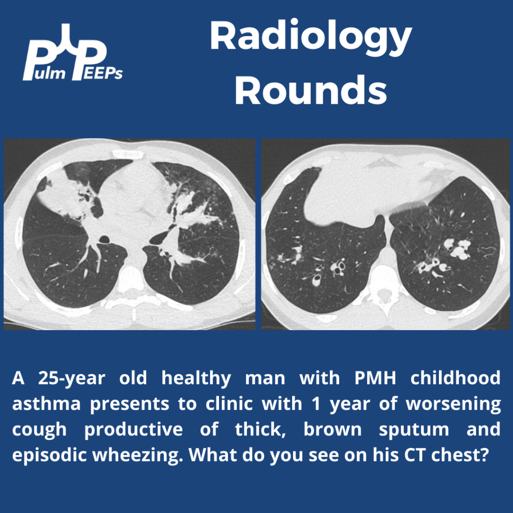
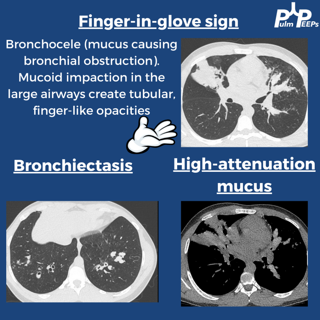
33 year old woman who came to the emergency department with acute onset of shortness of breath. She states that she had been in her normal state of health until this morning when she developed shortness of breath at rest, and chest pain. She does report a non-productive cough over the last few weeks which she feels may be contributing to her chest pain. She does report a history of asthma during childhood but without any exacerbations or maintenance therapies needed during her adulthood. She does report wheezing when she is sick with a cold but this is infrequent. The ED team sent off an initial work-up including a D-Dimer which was elevated, and she underwent a CTA of the chest for concern for possible PE. On the CT scan, there was no PE but the radiologist did call a “2 mm indeterminate right upper lobe pulmonary nodule.”
Case 2:
We have a 67-year-old male with a past medical history of ischemic cardiomyopathy, chronic systolic heart failure (LVEF 10-15%), s/p AICD, diabetes mellitus type 2, hyperlipidemia, hypertension, chronic kidney disease stage III, prostate cancer s/p seed implantation that was over 15 years ago who presented with acute decompensation of his heart failure and cardiogenic shock. He was successfully managed for that and is now being worked up by advanced HF and as a part of that workup got a chest CT, which found a RUL 6 mm nodule.
Case 3:
We have a 66-year-old male with a past medical history of HTN and drug abuse who presented to the ED with acute SOB, likely a COPD exacerbation. He was given bronchodilator and steroids as well as being started on Bipap. He eventually was able to be weaned off Bipap and was able to tolerate nasal cannula. As a part of his initial work up, the patient underwent CT scan for possible PE which demonstrated a new LUL spiculated nodule that is 1.3cm that is new since 2019.
Key Learning Points
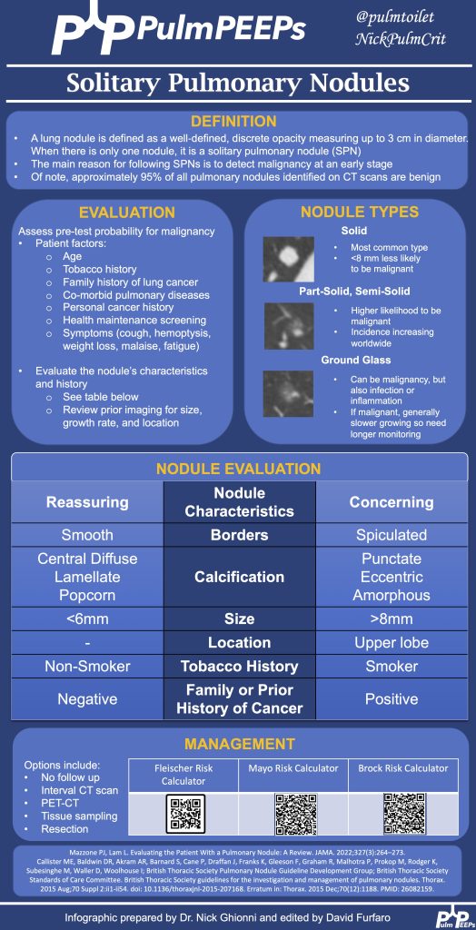
Approaching Pulmonary Nodules:
A structured approach is essential due to the complexities of diagnosing pulmonary nodules.
Patient history, including risk factors, past interventions, and imaging, plays a vital role.
Nodules’ appearance, such as location, shape, or characteristics like calcification or spiculation, can provide diagnostic clues.
The nodules history on serial imaging is a key predictive risk factor for determining the likelihood that the nodule represents cancer
Tools like the Mayo Risk Calculator and Fleishner Society guidelines assist in risk assessment and guidance.
It’s essential to assess patient risk, and nodule risk, and prioritize patient concerns and education. Periodic monitoring or follow-up might be necessary based on the nodule’s risk and size.
A multidisciplinary approach involving various specialists ensures comprehensive care.
Key Discussion Points:
PET Scans:
Useful in gauging a nodule or tumor’s metabolic activity.
Large, hypermetabolic nodules are suspicious.
Not every positive PET result means malignancy; other causes like inflammation or scars can produce positive results.
Evaluating Nodules:
Consideration of nodule size, characteristics, patient history, and risk calculators is crucial.
Tumor boards provide a collaborative expertise approach.
Tissue Sampling & Testing:
The method of tissue sampling depends on resources and expertise.
CT-guided biopsy offers a high diagnostic yield but with a risk of pneumothorax.
Bronchoscopic biopsy provides a lower diagnostic yield than CT-guided biopsy but has a significantly reduced risk of complications.
Advanced diseases now often require molecular testing on tissue samples.
Ground Glass Nodules:
Different from solid nodules due to their slow growth rate.
Monitoring is crucial due to the potential for transformations raising cancer suspicions.
The approach for ground glass nodules typically involves more extended monitoring intervals than for solid nodules.
Holistic Evaluation:
Consider the nodule’s characteristics, the patient’s history, and clinical intuition.
Individualized patient assessment is as vital as evidence-based guidelines and clinical expertise.
See the infographic for a summary of key learning points:
References and further reading
Loverdos K, Fotiadis A, Kontogianni C, Iliopoulou M, Gaga M. Lung nodules: A comprehensive review on current approach and management. Ann Thorac Med. 2019 Oct-Dec;14(4):226-238. doi: 10.4103/atm.ATM_110_19. PMID: 31620206; PMCID: PMC6784443.
Mazzone PJ, Lam L. Evaluating the Patient With a Pulmonary Nodule: A Review. JAMA. 2022 Jan 18;327(3):264-273. doi: 10.1001/jama.2021.24287. PMID: 35040882.
MacMahon H, Naidich DP, Goo JM, Lee KS, Leung ANC, Mayo JR, Mehta AC, Ohno Y, Powell CA, Prokop M, Rubin GD, Schaefer-Prokop CM, Travis WD, Van Schil PE, Bankier AA. Guidelines for Management of Incidental Pulmonary Nodules Detected on CT Images: From the Fleischner Society 2017. Radiology. 2017 Jul;284(1):228-243. doi: 10.1148/radiol.2017161659. Epub 2017 Feb 23. PMID: 28240562.
Wahidi MM, Govert JA, Goudar RK, Gould MK, McCrory DC; American College of Chest Physicians. Evidence for the treatment of patients with pulmonary nodules: when is it lung cancer?: ACCP evidence-based clinical practice guidelines (2nd edition). Chest. 2007 Sep;132(3 Suppl):94S-107S. doi: 10.1378/chest.07-1352. PMID: 17873163.
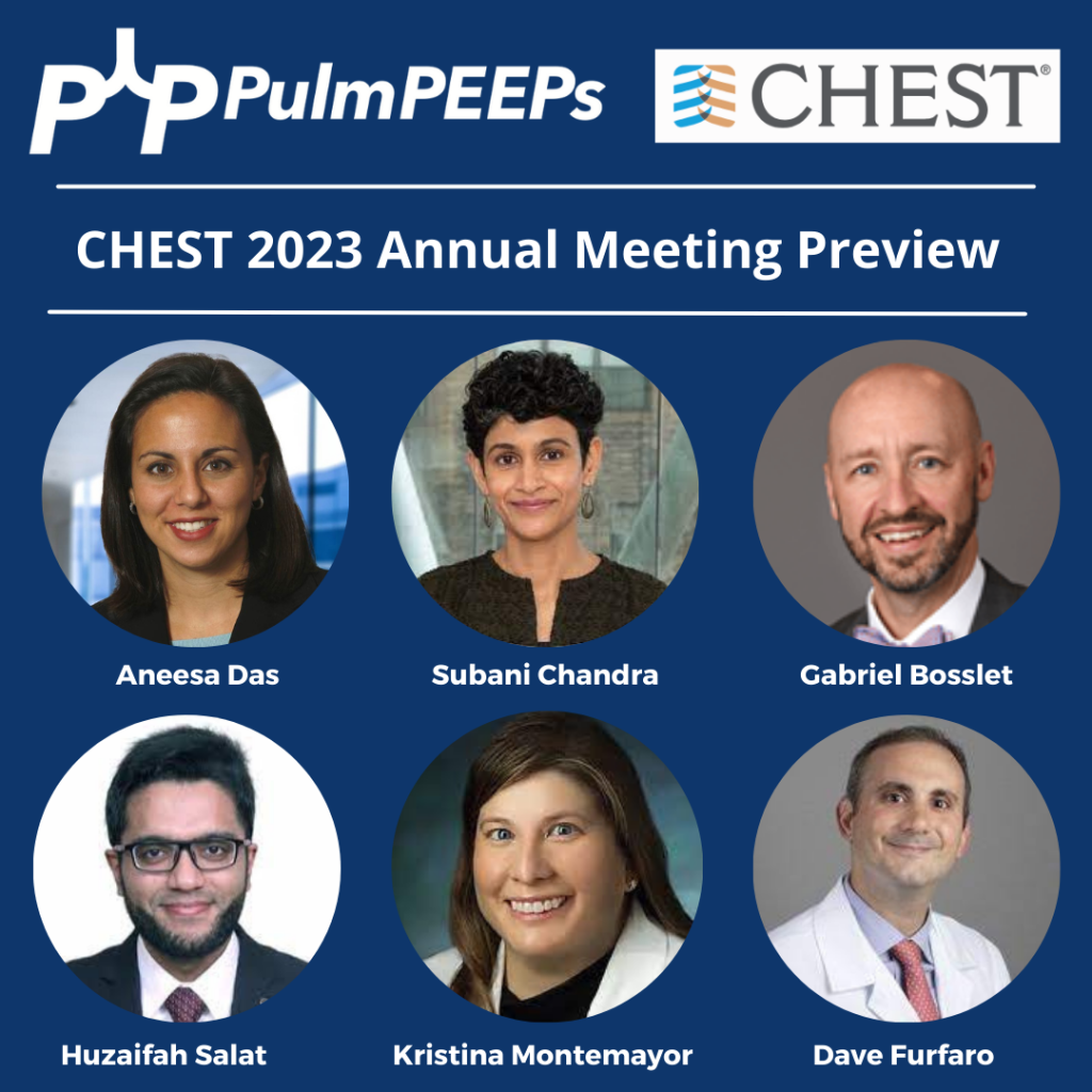
We are thrilled today to be previewing CHEST 2023! The Annual Meeting is taking place October 8th – 11th in Honolulu, Hawaii, and we are joined today by CHEST enthusiasts and the past, present, and future conference chairs. Listen now to hear what is in store for you next month in Hawaii, to plan your conference experience, and find out what sessions are can’t-miss!
Meet Our Guests
Aneesa Das is a Professor of Medicine at The Ohio State University Wexner Medical Center. She is the Assistant Director of the OSU Sleep Program and the Director of the Portable Sleep Testing Program. She was the Vice-Chair of the CHEST 2022 Scientific Programming Committee, and the Chair for 2023
Subani Chandra is an Associate Professor at Columbia University. She is the Vice Chair of Medicine for Education, and the internal medicine residency program director. She was the chair of the CHEST Scientific Program Committee for CHEST in 2022 and joined us when we came to you live from Nashville last year. Subani is currently the Chair for the Training and Transitions Committee for CHEST.
Gabe Bosslet is a Professor of Clinical Medicine in the Department of Pulmonary, Critical Care, Sleep and Occupational Medicine at Indiana University. He is the Assistant Dean for Faculty Affairs and Professional Development at IU. He is the current Vice Chair of the CHEST 2023 Scientific Programming Committee and the Chair Elect for CHEST 2024.
Huzaifah Salat is a budding clinician educator who is currently working as a consultant pulmonologist and intensivist at Advocate Aurora Health in Wisconsin. He recently completed his Pulmonary and Critical Care Fellowship at the University of Oklahoma Health Sciences Center. He has worked with Pulm PEEPs before on some fantastic Tweetorials.
CHEST’s Local Efforts and Initiatives to Support Survivors of the Maui Wildfires
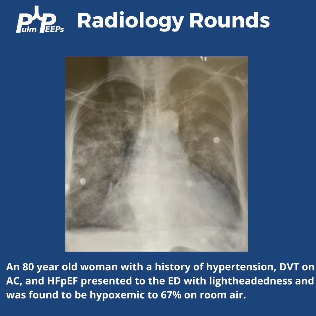
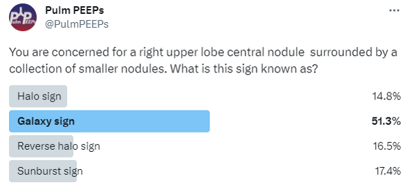
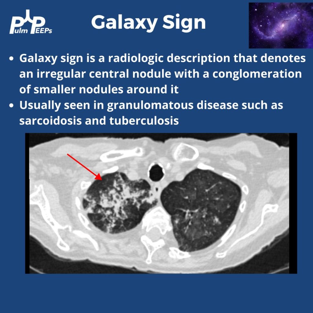
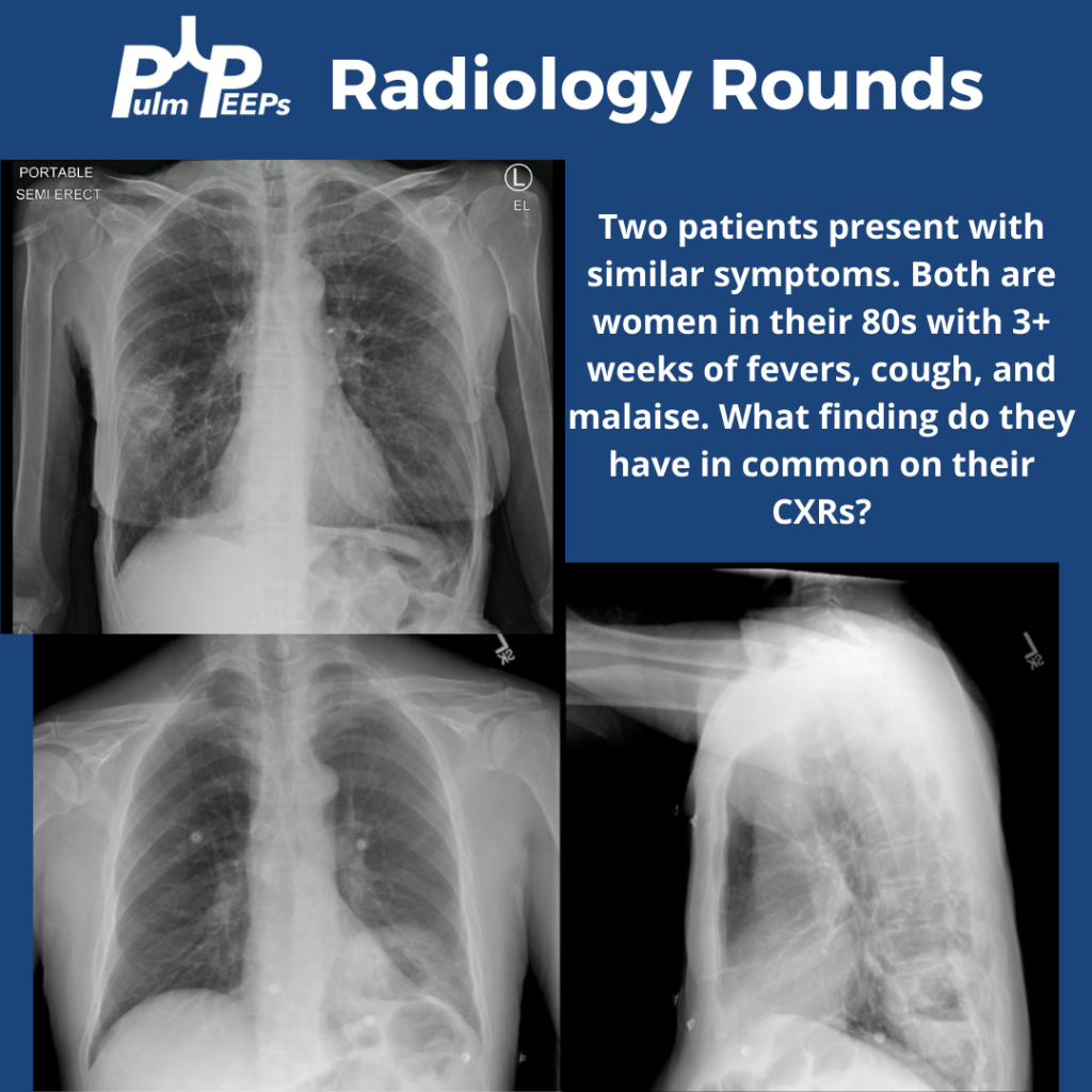
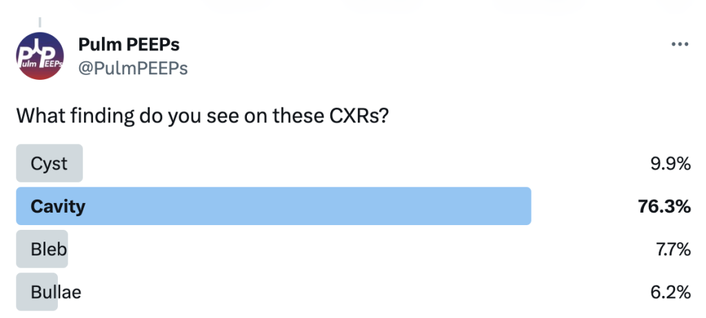
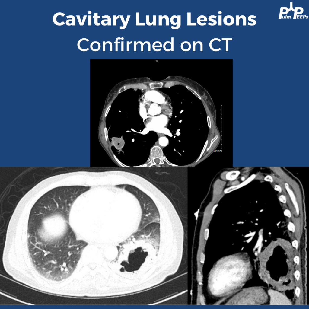
Tuesday is time for another #RadiologyRounds! Time for some CXR reading and a differential diagnosis mnemonic Two women presented to the hospital with similar presentations. They are both in their 80s with multiple weeks of cough, fever, and fatigue. Here are the CXRs
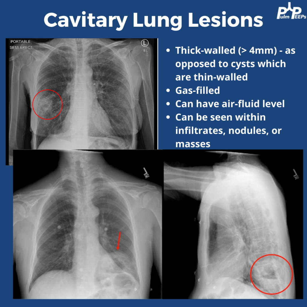
The CXRs both showed cavities. They are thick-walled (>4mm) and gas-filled. Cavitary lung lesions are seen within infiltrates, nodules, or masses. There can be an air-fluid level within the cavity. Cysts have thinner walls. The findings were confirmed on CT scan
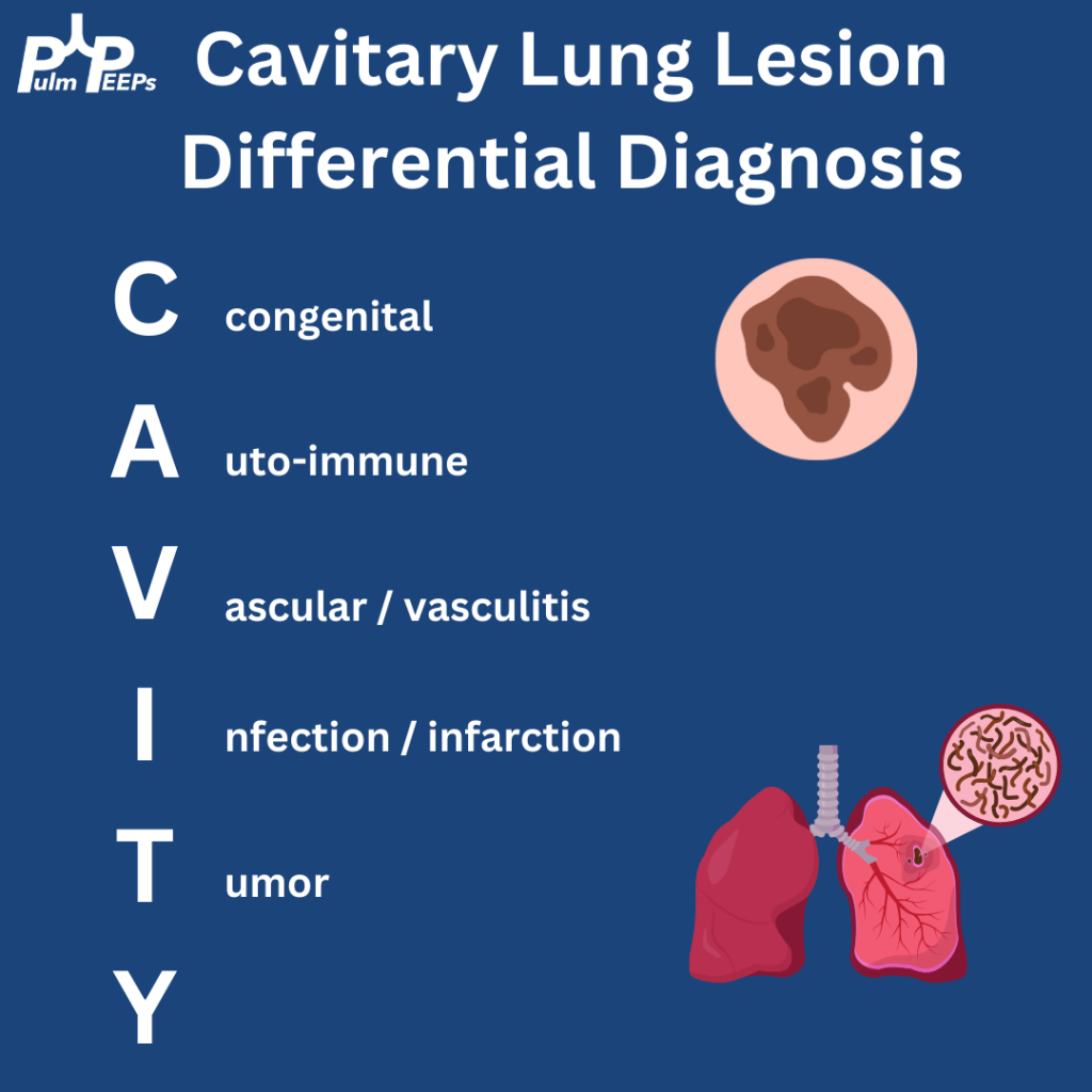
Cavitary lung lesions can have a broad differential so it is helpful to have a systematic approach. To make it easy, when you see this just remember: CAVITY
Bonus points to anyone who can fill in the Y
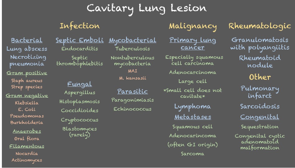
Both patients were ultimately diagnosed with pulmonary abscesses which improved with prolonged courses of antibiotics with anaerobic and gram-negative coverage.
We’re excited to be back with another Fellows’ Case Files. Today, we’re visiting the University of Pittsburgh to meet a fantastic fellow and a dedicated educator, and to hear about a fascinating case. Let us know if you’ve ever had a similar case, and share your interesting cases with us!
Meet Our Guests
Rachel Wojcik obtained her B.S. in Biology from Mercyhurst University and a Master’s in Liberal Studies from the University of Denver in Global Affairs with a focus on Healthcare. She completed her MD at the University of Colorado before completing her residency and chief resident year at the University of Pittsburgh and has continued her training at Pitt for PCCM fellowship.
Dr. Stephanie Maximous is an Assistant Professor of Medicine at the University of Pittsburgh School of Medicine and is the Clinical Education APD for the Pulmonary and Critical Care Fellowship program. She completed her fellowship at Pitt in addition to obtaining a Master’s Degree in Medical Education there. She teaches in and directs courses throughout the medical school, residency, and fellowship and was recently awarded the 2023 Outstanding Subspecialty Teaching Attending Award from the housestaff.
Case Presentation
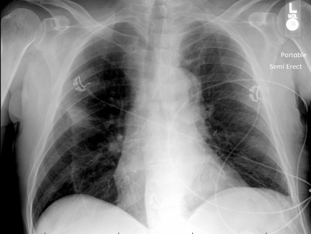
Patient: A 70-year-old male with a history of idiopathic thrombocytopenia on chronic prednisone and a history of tobacco use disorder.
Presentation: Came to the hospital with 2-3 days of right-sided weakness and slurred speech.
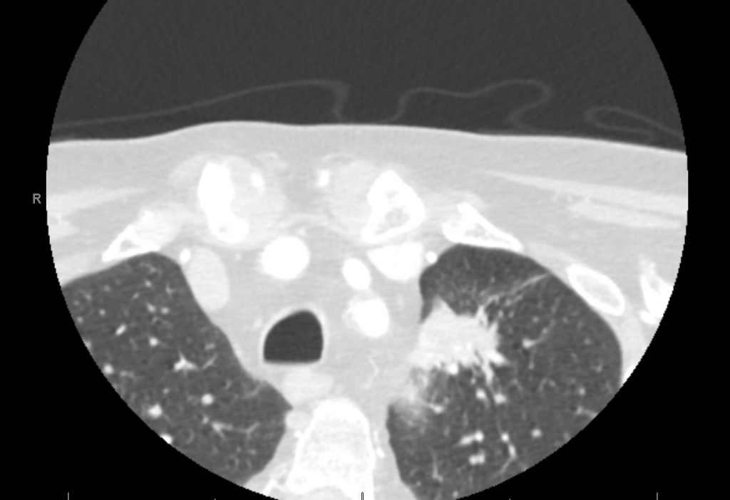
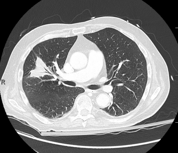
Findings: MRI showed a moderate-sized left pontine stroke. A CT angiogram of the neck showed no evidence of an occlusion, but a spiculated two-centimeter nodule at the apex of the left lung was found.
Additional Information: He requires a walker for mobility and needs help with activities like taking a shower and dressing. He had an unintentional 20-pound weight loss over six months, increased fatigue, and malaise.
Previous Investigations: A chest x-ray ordered two months prior by his hematologist was unremarkable, and a CT of the abdomen and pelvis showed no masses.
Key Learning Points
Bronchoscopy in Decision Making:
The decision to perform bronchoscopy in patients depends on a myriad of factors, including the location of any lesions, accessibility, potential risks, and the potential diagnostic yield.
Fiber optic bronchoscopy with BAL can rule out infections, and if no diagnosis is reached, more invasive methods like surgical biopsy might be necessary.
Consider the location of consolidated masses; navigational bronchoscopy might be needed for lesions without a clear airway leading into them.
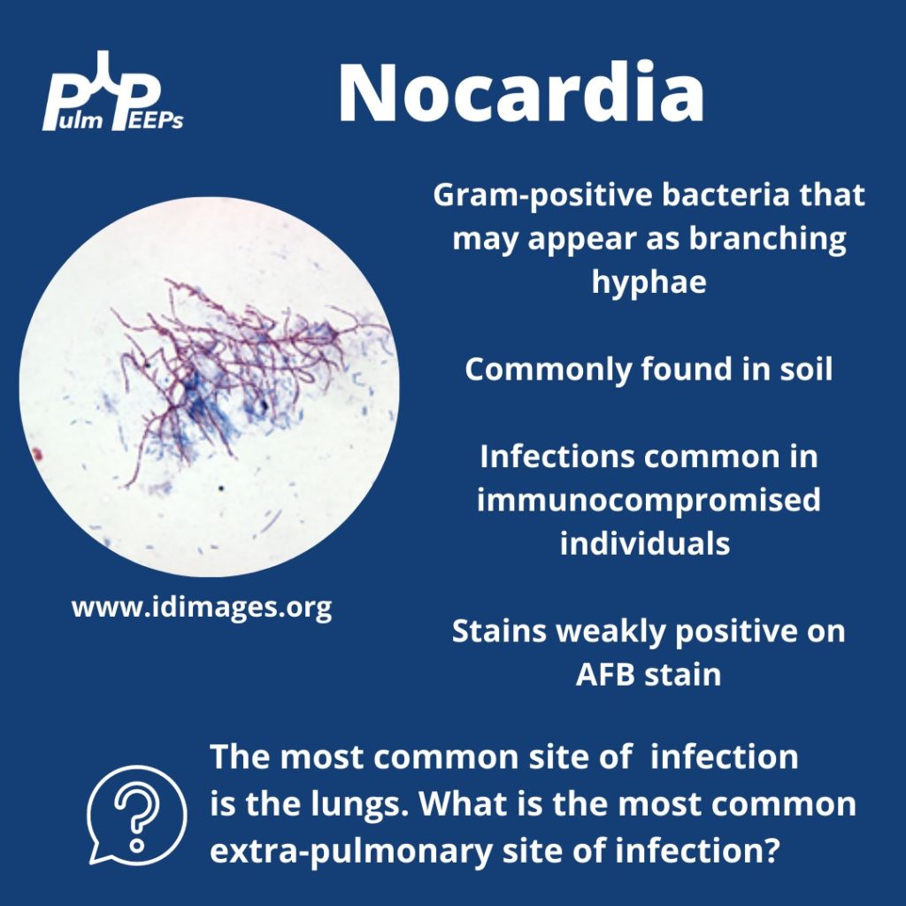
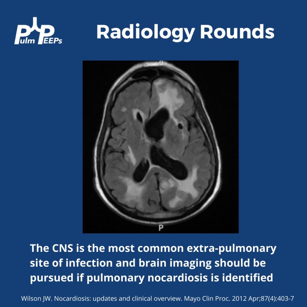
Nocardia Insights:
Nocardia is a gram-positive bacterium that stains weakly acid-fast.
It can be found in soil and certain water sources and can infect through the skin or by inhalation.
Two-thirds of patients with Nocardia are immunocompromised.
The dosage of Bactrim given for PJP prophylaxis doesn’t prevent Nocardia infections in immunocompromised individuals.
While the lungs are the most common infection site, Nocardia can manifest elsewhere, like the skin or CNS.
Treatment Approach:
Bactrim is the mainstay of treatment for Nocardia. If someone is allergic, desensitizing them can be crucial.
IV induction phases vary in length depending on the severity of the disease.
The overall treatment duration is protracted to prevent relapse.
Takeaway Points:
Bactrim for PJP prophylaxis doesn’t necessarily prevent Nocardia infections in immunocompromised individuals.
If someone is allergic to Bactrim, consider desensitizing them due to its importance in treating Nocardia.