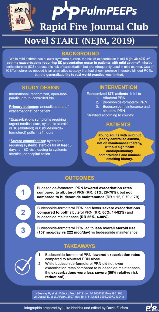
We are excited to bring an a dedicated episode all about inhalers. We know there are many type of inhalers, formulations and techniques that are needed for successful use and we cover them all. Take a listen today!
Meet our Guests
Amber Lanae Martirosov is an Associate Clinical Professor at Wayne State University and is an Ambulatory Care Pharmacy Specialist in Pulmonary at Henry Ford Health in Detroit, Michigan. Amber’s specific interests include appropriate inhaler use, medication access, ILD and advocating for pharmacy collaborations.
Nick Ghionni is a first year attending at the MedStar Baltimore Hospital System. He is fresh out of PCCM fellowship at MedStar Washington Hospital Center. He completed his Internal Medicine residency at Mercy Catholic Medical Center and his specific interests include mechanical ventilation, POCUS, and medical education. Nick is our newest member of the PulmPEEPs team and serves as an Associate Editor.
Device Overview
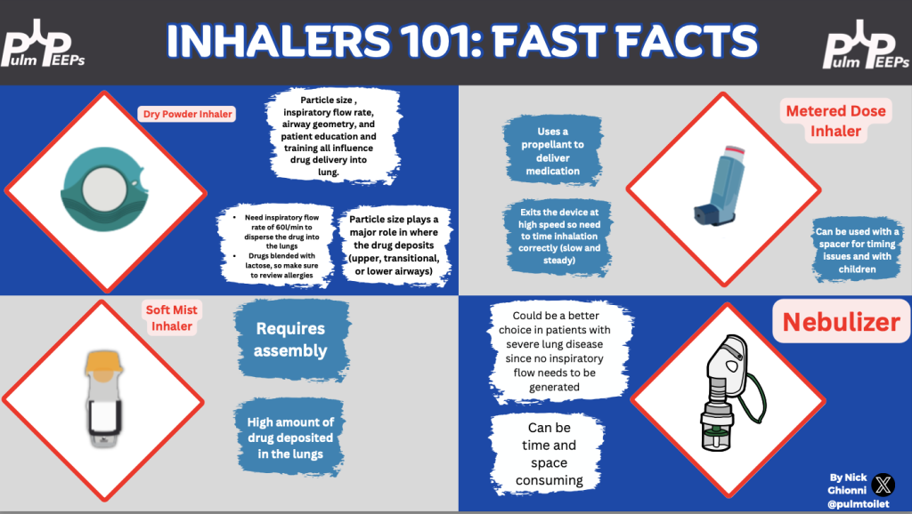
1. Metered dose inhaler (MDI): delivers a dose of medication when you press on the canister. 2. Dry powder inhaler (DPI): delivers powered medication with each inhalation. 3. Soft mist inhaler (SMI): which sprays a dose of medication when pressed
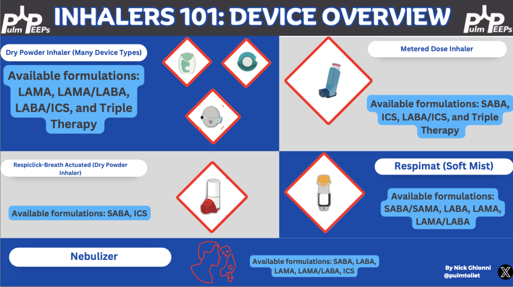
Inhaler Charts
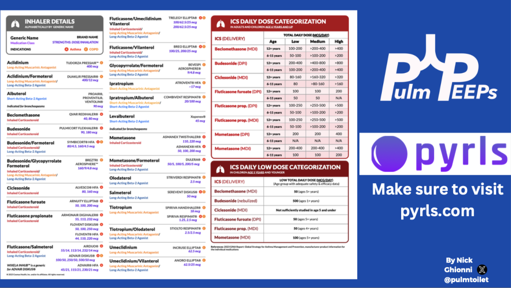
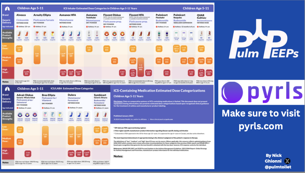
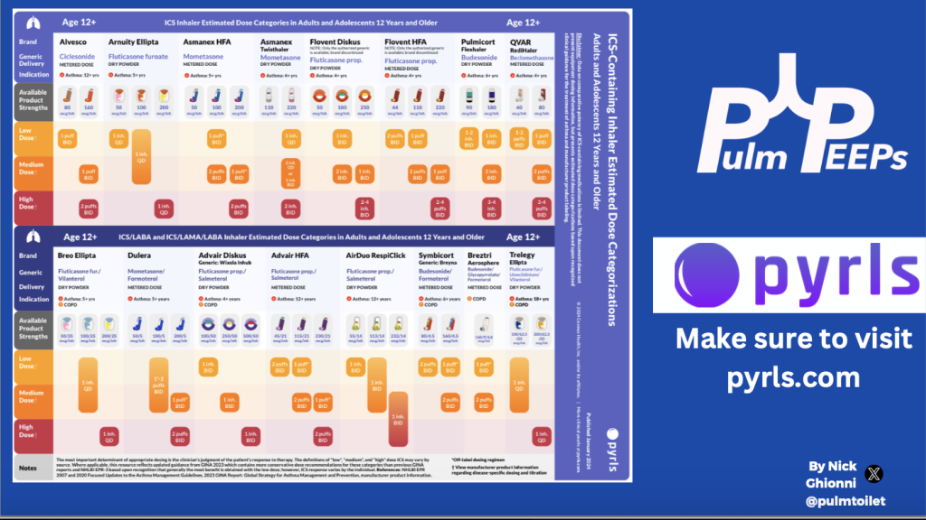
We partnered with Pyrls to show common inhaler devices, formulations and dosing. You can create a free Pyrls account at pyrls.com or our app they can download an additional bundle/more awesome charts just like these totally free!

Additional Resources
References and Further Reading
Brand P, Hederer B, Austen G, Dewberry H, Meyer T. Higher lung deposition with Respimat Soft Mist inhaler than HFA-MDI in COPD patients with poor technique. Int J Chron Obstruct Pulmon Dis. 2008;3(4):763-70. PMID: 19281091; PMCID: PMC2650591.
Levy ML, Carroll W, Izquierdo Alonso JL, Keller C, Lavorini F, Lehtimäki L. Understanding Dry Powder Inhalers: Key Technical and Patient Preference Attributes. Adv Ther. 2019 Oct;36(10):2547-2557. doi: 10.1007/s12325-019-01066-6. Epub 2019 Sep 2. PMID: 31478131; PMCID: PMC6822825.
Jindal S K, Pandey K K, Bose P P. Dry powder inhalers: Particle size and patient-satisfaction. Indian J Respir Care 2021;10:14-8
Spitzer WO, Suissa S, Ernst P, Horwitz RI, Habbick B, Cockcroft D, Boivin JF, McNutt M, Buist AS, Rebuck AS. The use of beta-agonists and the risk of death and near death from asthma. N Engl J Med. 1992 Feb 20;326(8):501-6. doi: 10.1056/NEJM199202203260801. PMID: 1346340.
Chang, YL., Ko, HK., Lu, MS. et al. Independent risk factors for death in patients admitted for asthma exacerbation in Taiwan. npj Prim. Care Respir. Med. 30, 7 (2020). https://doi.org/10.1038/s41533-020-0164-4
Podcast: Play in new window | Download
Subscribe: Apple Podcasts | Spotify | Amazon Music | Android | iHeartRadio | Podcast Index | RSS