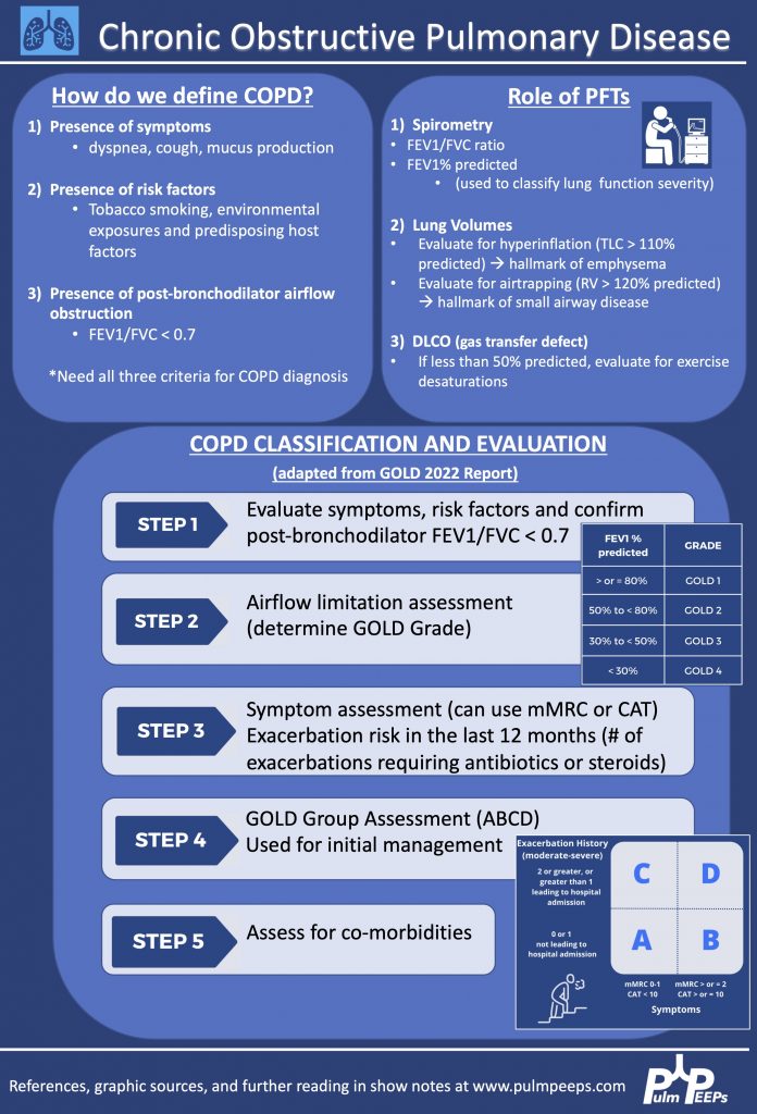
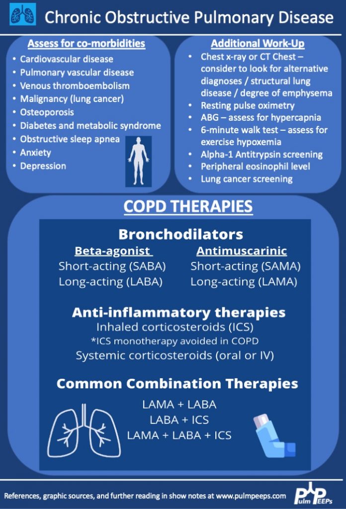
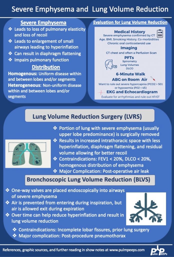
Evaluating a patient with suspected interstitial lung disease is a challenge for all pulmonary clinicians from medical students to attending physicians. This week’s Radiology Rounds covers some findings seen in one classic pattern. Make sure to listen to our show next week for more about ILD diagnosis!
Dave Furfaro, Kristina Montemayor, and Ansa Razzaq are back to tackle another pulmonary case! Listen in and solve the case yourself, and we’ll share some diagnostic pearls along the way. Let us know any additional thoughts on Twitter.
Patient Presentation
The is patient is 57-year-old man with hypertension and asthma who presents with dyspnea and left-sided pleuritic chest pain for 3 weeks. He was in his usual state of health until 3 weeks prior to admission, when he developed dyspnea and sharp left-sided chest pain that worsens with deep breathing. His symptoms are accompanied by unintentional 30-pound weight loss over the past several months as well as an intermittent cough that is nonproductive.
On physical exam, he is mildly tachypneic and saturating well on room air with otherwise normal vital signs. He has decreased breath sounds at the right lung base.
Initial labs
Key Learning Points
**Spoilers Ahead** If you want to think through the case on your own we advise listening to the episode first before looking at the infographics below
Physical Exam Pearls
Reasons for decreased breath sounds on physical exam
1. Increased thickness of chest wall
2. Reduced airflow to part of the lung
3. Overinflation to part of the lung
4. Something between the lung and chest wall — air or fluid
Determine bradypnea and tachypnea quickly by matching your breahting rate to the patient’s respiratory rate
1. Is there a compatible presentation (imaging, physical exam)
2. Detection of non-necrotizing granulomatous inflammation in one or more tissue samples
3. Exclusion of other disease that may present similarily
Pulmonary stages of sarcoidosis:
Key to remember that patients don’t always progress through these stages. The system is useful for prognosticating and determining treatment based on the risk for disease progression.
Patients with stage 1, and even many with stage 2, often don’t require treatment
The first-line agent is oral glucocorticoids and the typical starting dose is prednisone 20 – 40 mg by mouth daily. The patient should be evaluated closely, and ideally, this dose can be tapered starting at about 4 – 6 weeks. Following this, the prednisone dose is tapered slowly over 6 months – 1 year while monitoring for symptom recurrence.
Second-line steroid-sparing agents are methotrexate, azathioprine, or mycophenolate. These are often used if the patient relapses, or is on more then 10mg daily for 3 months after the initial taper with intolerance of steroids
This week for radiology rounds we’re looking at some classic imaging signs of lobar collapse. Take a look, respond to our polls on Twitter, and make sure to subscribe to our podcast to get all of the Pulm PEEPs content!
We are excited to bring you another episode in our Pulm PEEPs Top Consults series! Kristina Montemayor and David Furfaro, are joined by Sandy Zaeh to discuss the assessment and management of a patient with a severe asthma exacerbation. We’ll follow a consult patient from the emergency department to the ICU, and cover everything from the physiology of pulsus paradoxus in asthma to how to manage the ventilator in status asthmaticus. Listen today and please send any questions our way on Twitter @pulmPEEPS.
Meet Our Guests
Sandy Zaeh is an Instructor of Medicine and Pulmonary & Critical Care Medicine physician at Yale School of Medicine.
The first Radiology Rounds of 2022! Follow the thread on Twitter to answer our live polls, and be sure to subscribe to the show wherever you listen to podcasts. This week we look at a distinctive, although not specific, pattern and a rare and fascinating diagnosis.
This week on Pulm PEEPs, Dave Furfaro and Kristina Montemayor are joined by experts in the field of critical care medicine and ARDS to discuss all things PEEP! Drs. Roy Brower, Sarina Sahetya, Todd Rice, and Elias Baedorf-Kassis discuss everything ranging from PEEP basics to their approach to optimizing PEEP in patients with ARDS.
Meet Our Guests
Roy Brower is a Professor of Medicine at Johns Hopkins where he served as the MICU director for over 33 years, and he has been one of the pioneers for lung-protective ventilation for patients with ARDS.
Elias Baedorf-Kassis is an Assistant Professor of Medicine at Beth Israel Deaconess Medical Center and Harvard Medical School. He is the Medical Director of Respiratory Care at BIDMC, and helps lead the VV-ECMO program.
Todd Rice is an Associate Profess of Medicine in the Division of Allergy, Pulmonary, and Critical Care Medicine at Vanderbilt University and Vice President for Clinical Trial Innovation and Operations in the Vanderbilt Institute for Clinical and Translational Research.
Sarina Sahetya is an Assistant Professor of Medicine at Johns Hopkins Hospital and does research in the diagnosis and treatment of ARDS.
Key Learning Points
Driving Pressure figure from Amato et al. 2015. Stress index figure from Hess 2014.
The plateau pressure can be measured on the ventilator with an inspiratory hold maneuver
Extrinsic PEEP is applied by the ventiilator, while intrinsic PEEP, or auto-PEEP, occurs when there is incomplete emptying of the lungs due to inadequate time for exhalation. This often happens with obstructive lung disease. Intrinsic PEEP can be measured on the ventilator with an end-expiratory hold maneuver
We utilize PEEP in all intubated patients to minimize atelectasis. When patients are supine, the heart moves back 2 cm and the diaphragm raises by 2 cm, so often the left lower lobe of the lung is compressed and there is atelectasis there. This is often seen on CXR:
Today we’re bringing you a special edition of Radiology Rounds complete with classic imaging, and some key critical care and ventilator physiology. This case is a perfect lead-in for next week’s Pulm PEEPs Roundtable on PEEP titration, so make sure to tune in!
How would you best describe the imaging findings?
There are bilateral, diffuse alveolar infiltrates noted on imaging with evidence of an air bronchogram on the CT image.
The patient develops worsening hypoxemia requiring mechanical intubation. The patient has multifocal pneumonia and requires intubation. ABG is performed and the calculated PaO2:FIO2 ratio is 150. How would you describe the severity of ARDS?
This patient has moderate ARDS based on a PaO2:FIO2 ratio that is between 100 and 200. The patient’s initial ventilator settings on volume control are:
Based on these parameters, we can also calculate the driving pressure. Driving pressure is calculated by using Pplat-PEEP. In this case, Pplat (30)-PEEP (10), would give a driving pressure of 20.
The Pulm PEEPs are joined again by Natalie West to discuss a patient who presented with a chronic, productive cough. Listen in today as we work through our differential diagnosis, interpret basic pulmonary testing, and share our clinical reasoning along the way. We have some fantastic diagnostic and treatment teaching points, so once you’ve solved the case check out the takeaways and infographics below. Please let us know any additional insights you have on Twitter!
Patient Presentation
A 50-year-old woman, who is a never smoker, with a past medical history of recurrent pancreatitis presents to the pulmonary clinic with a chronic, productive cough. Her cough has been present for 3 years and has increased in frequency to now being present daily. In the last three months, the cough has also worsened and is productive of small amounts of yellow to green sputum. She has a history of chronic post-nasal drip and sinus infections, and uses intranasal steroids, but has not noted changes in these symptoms. There is no significant family history of pulmonary disease, and an exposure history review of symptoms is negative.
On physical exam, she was a thin woman who appeared her stated age and was breathing comfortably on room air. Her exam was notable for mild expiratory wheezing, primarily on auscultation of the right posterior lung field. She had no cyanosis, clubbing, evidence of volume overload, or abdominal tenderness.
Basic Spirometry Values
Chest X-ray
Representative Images from CT Scan
Key Learning Points
**Spoilers Ahead** If you want to think through the case on your own we advise listening to the episode first before looking at the infographics below.
Differential Diagnosis of Chronic Cough
Three most common causes: upper airway cough syndrome, GERD, cough variant asthma
Additional etiologies to consider: chronic bronchitis, post-infectious after a respiratory tract infection, bronchiectasis, ACE inhibitors, lung cancer, eosinophilic bronchitis, interstitial lung disease
Imaging Pearl
Evaluating Bronchiectasis
Making a New Diagnosis of Cystic Fibrosis in an Adult
Sweat testing
Sweat testing should be done in CF accredited center. Inform patients that there are no needles involved. Pilocarpine and electrical stimulation are applied to the arm or leg to stimulate the sweat gland, and then sweat is collected on filter paper, a gauze, or a plastic coil. From there, the amount of chloride in the sweat is calculated
Results
< 30 normal
31 – 60 indeterminate
> 60 is positive and Cystic Fibrosis is likely
What do you do with an Indeterminate test?
Patients with milder phenotypes of Cystic Fibrosis can have a normal or indeterminate sweat chloride level, and 10% of adults diagnosed with CF have a normal sweat chloride. If the sweat chloride test is indeterminate or normal, but suspicion is high for CF, then genetic testing for the whole array of mutations should be performed
Pulm PEEPs hosts, Kristina Montemayor and David Furfaro, bring our first episode in our Top Consults series. In this series, we will bring in experts to work through the most common pulmonary and critical care consults. Whether you are the consulting physician, or a pulmonologist responding to the page, these episodes are geared to give you all the information you need to care for your patients!
Today, we are joined by Chris Kapp and Matthew Schimmel, two interventional pulmonologists, to discuss hemoptysis. Chris and Matt will help us work through two hemoptysis consults, and together we’ll provide a framework for thinking about hemoptysis, outline some key components of the evaluation, and delve into treatment options.
Key Learning Points
Hemoptysis Evaluation
Hemoptysis Management
Life-Threatening or Large Volume Hemoptysis
Stabilize the patient! Make sure the airway is protected either by the patient coughing themselves, or intubation if needed. Provide hemodynamic support with IVF, blood products, and pressors if needed. If it is known which lung has the bleeding the patient can be positioned so the lung with the bleeding is down. This protects the non-bleeding lung.
Correct any bleeding diathesis If the patient is on anti-coagulation, or has any reversible bleeding diathesis, these should be corrected immediately to reduce further bleeding.
Localize the bleed If the patient is stable, they should undergo a CTA to localize the bleeding. If they are not stable to make it to a CT scan, a bronchoscopy should be performed.
Bronchoscopic treatment In addition to clearing blood from the airway, bronchoscopy can localize the bleeding. With available expertise, bronchoscopic treatments can be performed such as ice saline, topical epinephrine, or balloon tamponade to isolate the bleed.
Definitive therapy with arteriography and embolization Patients with life-threatening hemoptysis should ultimately undergo arteriography and embolization of any bleeding vessel. If this is not possible, then surgery can be needed in some cases.
A note on diffuse hemoptysis If there is not one distinct bleeding lesion, then localizing and treating the bleed becomes more difficult. For diffuse alveolar hemorrhage, evaluation should be performed for if it is primary, and due to an immunologic cause and capillaritis, or secondary to a systemic disease and / or bleeding diathesis. These investigations will guide available treatment options. Capillaritis from an immunologic cause, such as lupus or vasculitis, can be treated with systemic glucocorticoids and an additional immunosuppressive agent such as cyclophosphamide or rituximab.
Non-life-threatening or Small Volume Hemoptysis
Monitor for clinical worsening Patient’s should be monitored, either in the in-patient or out-patient setting, for increased volume or frequency of hemoptysis and for any clinical worsening, such as desaturations or decreased ability to clear the airway.
Correct any bleeding diathesis If the patient is on anti-coagulation, or has any reversible bleeding diathesis, these should be corrected immediately to reduce further bleeding. In pattients with non-life-threateneing hemoptysis this requires careful consideration of balancing the risk of bleeding vs the benefits for continuing anti-coagulation.
Evaluate for underlying cause Patient’s should undergo imaging and evaluation for the underlying cause of the hemoptysis. This may be evidence of an underlying infection, a pulmonary embolism, or new lung lesions making the patient at risk. If the source can’t be found on non-invasive imaging, and there is no clear systemic source such as an infection, a bronchoscopy is warranted. Any underlying cause should be treated and investigated further.
Inhaled Tranexamic Acid Nebulized tranexamic acid is well tolerated and can help resolve hemopytysis without invasive procedures.