Welcome to another episode in our Pulm PEEPs Fellows’ Case Files series! The purpose of this series is to highlight and amplify the incredible clinical work that is done by pulmonary and critical care fellows, share fascinating cases, and assemble a diverse network of pulmonary and critical care educators. Today we’re headed to Baylor College of Medicine to hear about a fascinating case. Tune in, let us know what you think on Twitter, and let us know if you have a great case to share!
Meet Our Guests
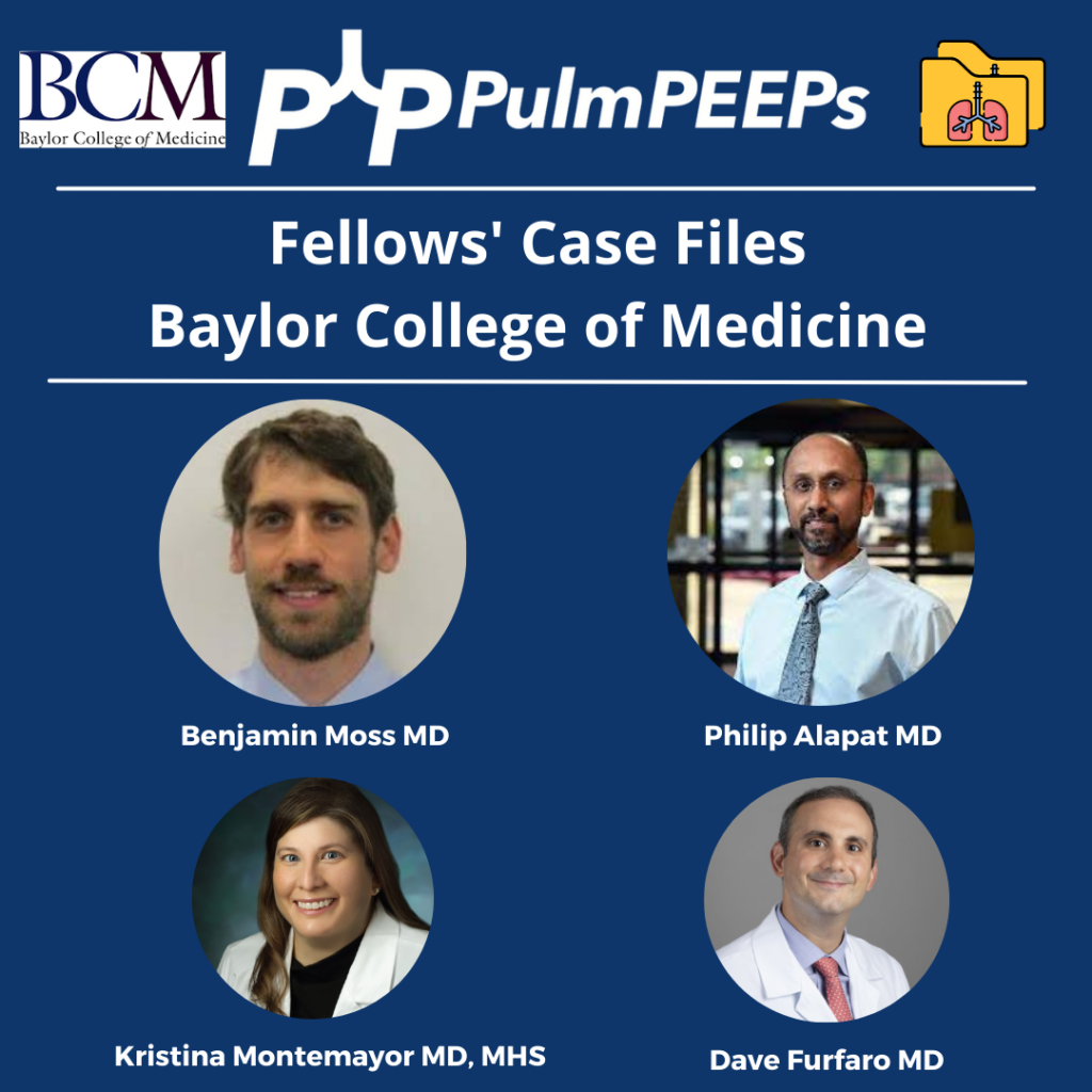
Benjamin Moss completed his internal medicine residency training at Baylor College of Medicine in Houston, Texas, and is currently a senior pulmonary and critical care fellow there.
Philip Alapat is an Assistant Professor of Medicine and the Program Director of the Pulmonary and Critical Care Fellowship at Baylor. He completed his residency and fellowship training all at Baylor.
Patient Presentation
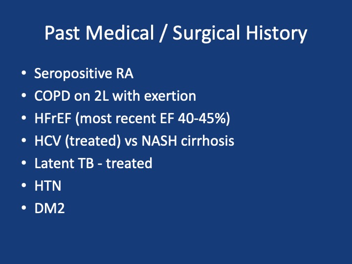
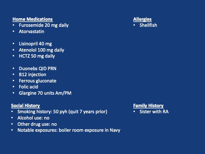
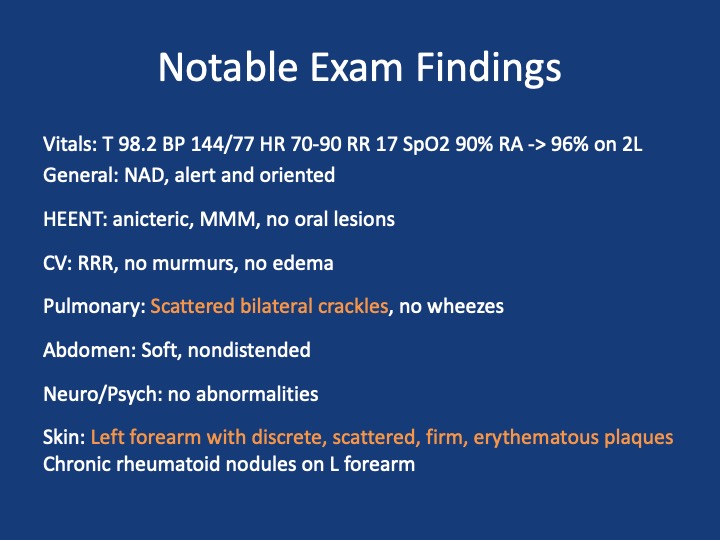
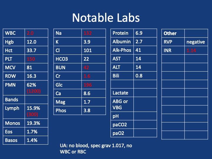
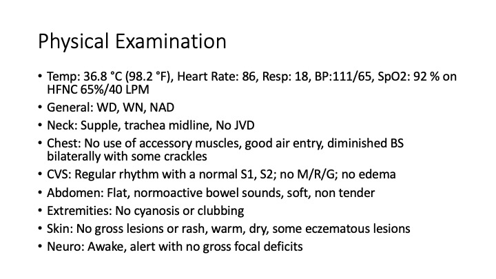
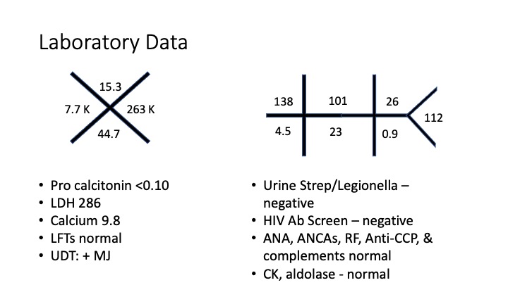
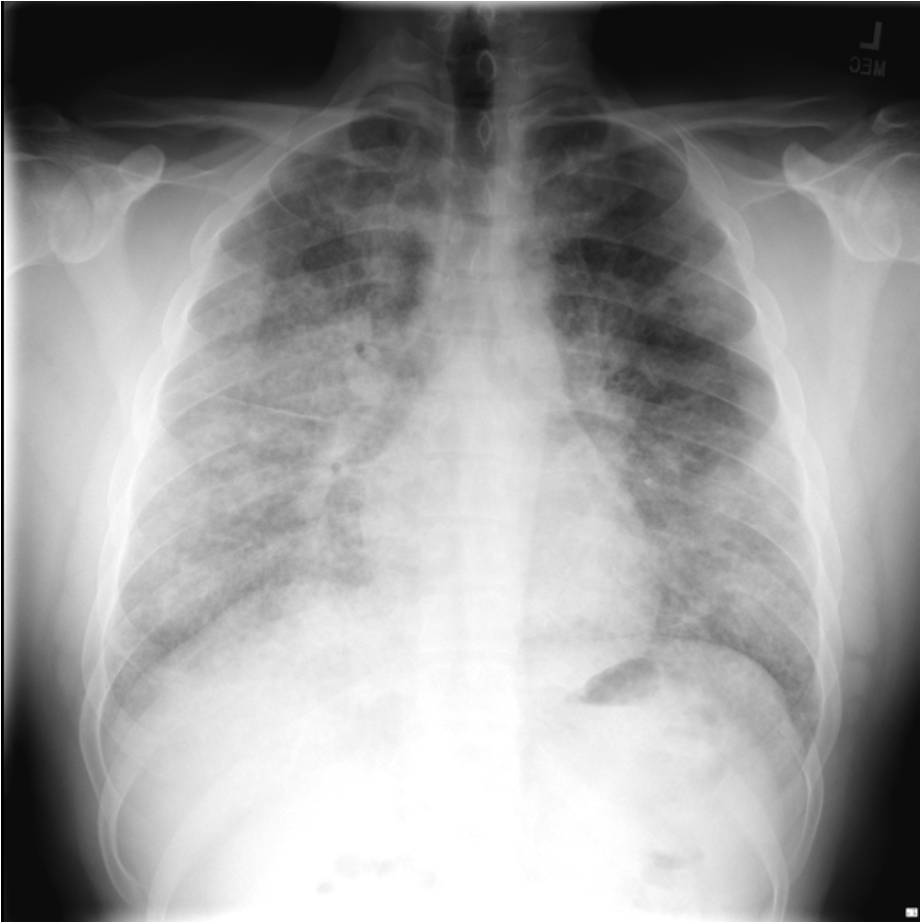
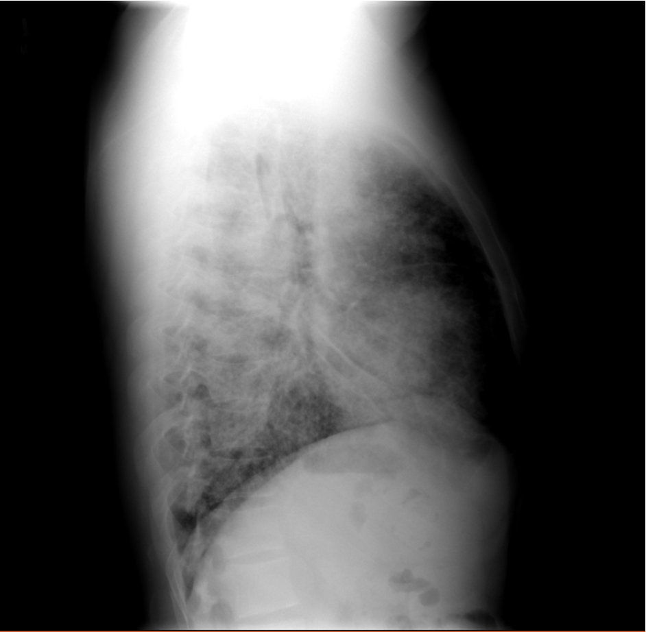
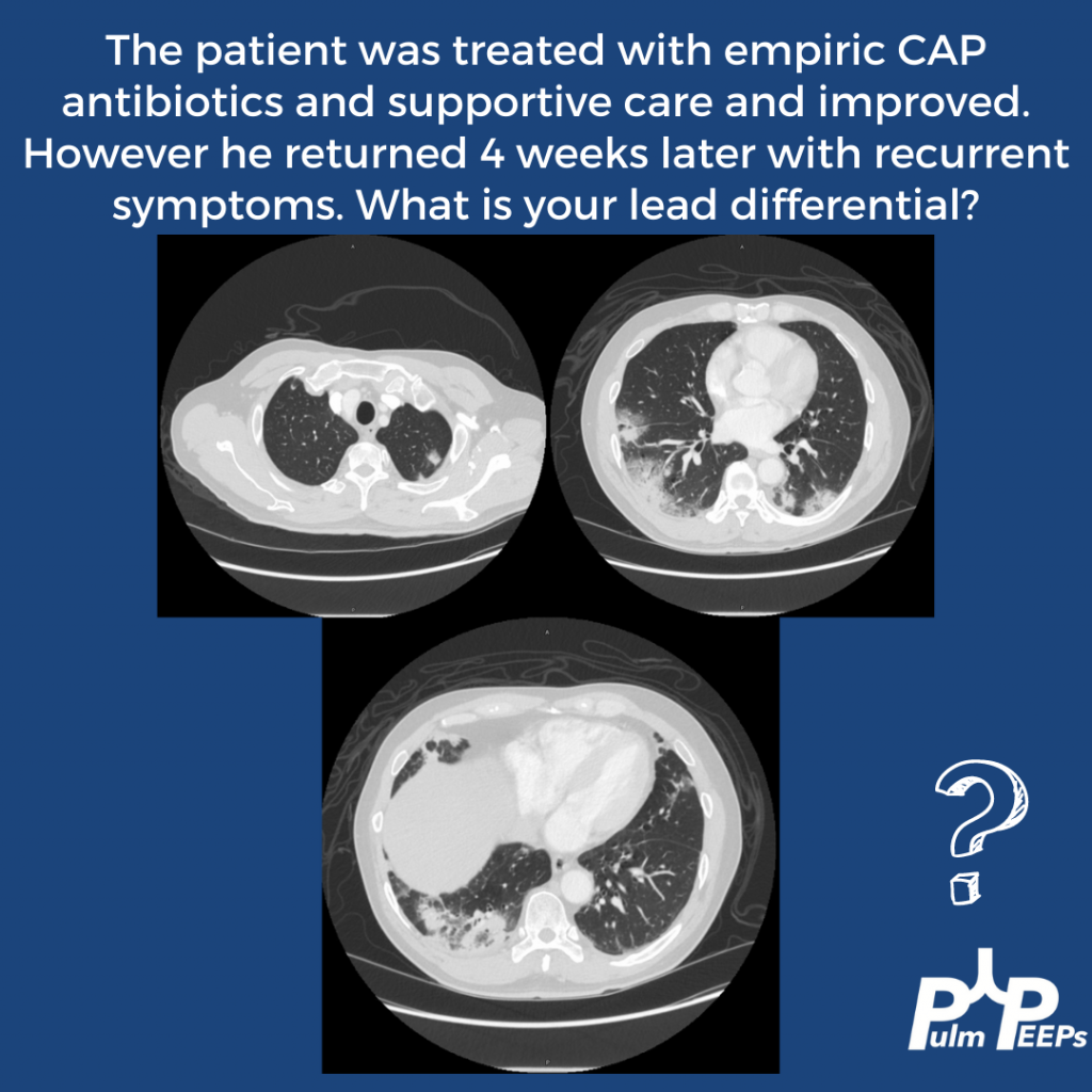
A 60s-year-old man with seropositive RA on Rituximab presents with dyspnea and cough, and overall “not feeling well.” For the past week, he has had malaise, body aches, and subjective fever. For the past 3 days, he has had acutely worsening dyspnea that is worse with exertion, but present at rest and a cough with scant sputum production. He had been on Methotrexate previously but within the last year developed pancytopenia and MTX was stopped and he was switched to adalimumab/Humira. His pancytopenias did not resolve, and he was ultimately diagnosed with Felty syndrome (a triad of RA, neutropenia, and splenomegaly) and switched to rituximab every 6 months with his last dose being 4 months ago. During the last week, he tried taking prednisone 10 mg a day but his symptoms did not improve.
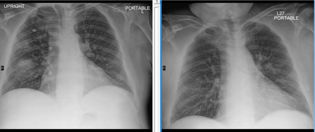
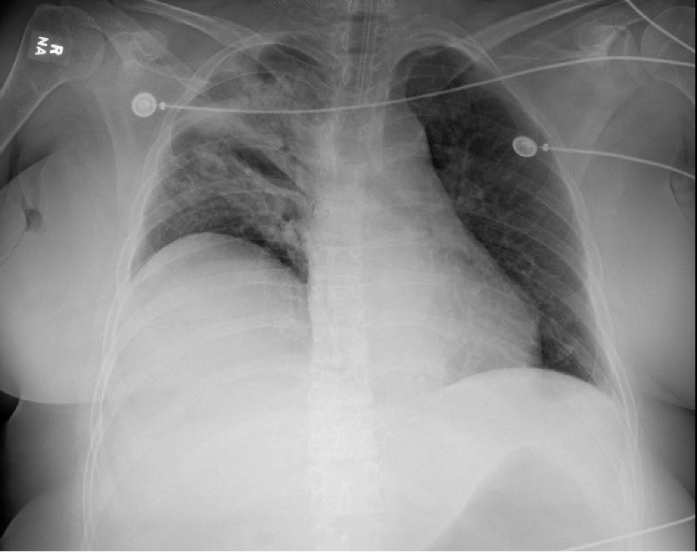
X-ray on presentation (L) X-ray 8 months ago (R)
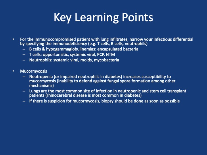
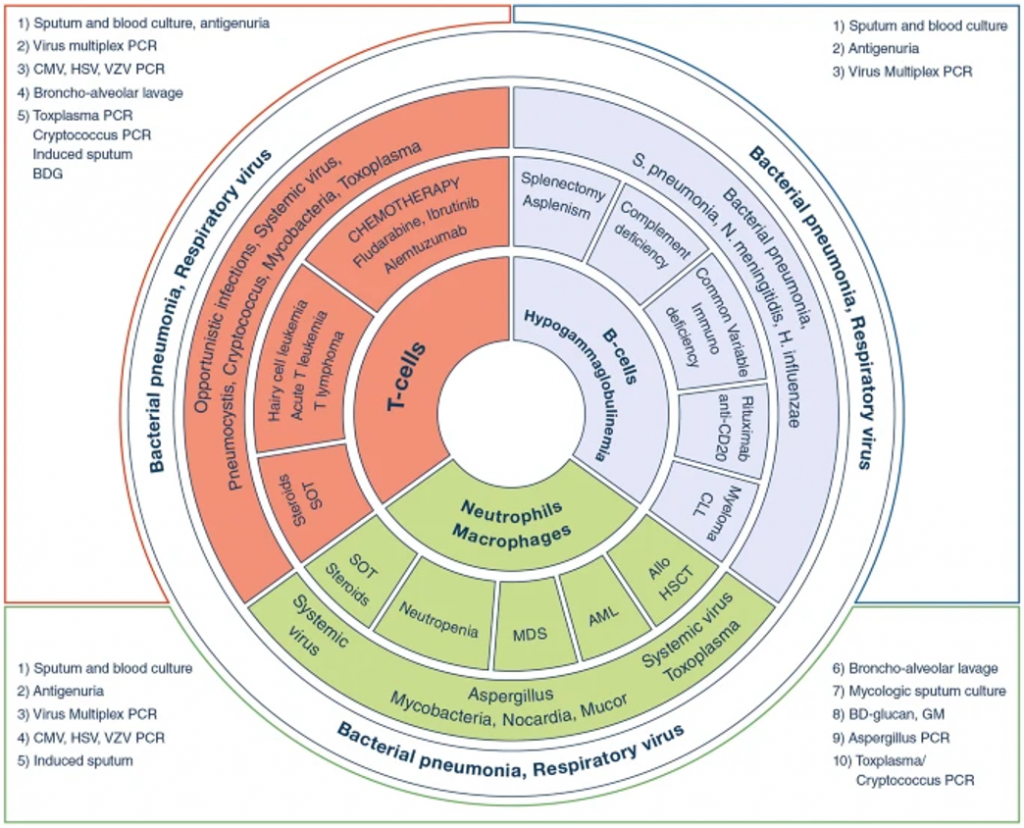
Key Learning Points
**Spoilers Ahead** If you want to think through the case on your own we advise listening to the episode first before looking at these points.
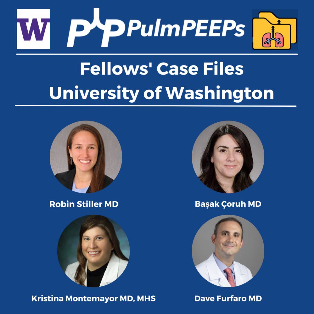
We’re very excited for the second episode in our Pulm PEEPs Fellows’ Case Files series! For a reminder, the purpose of this series is to highlight and amplify the incredible clinical work that is done by pulmonary and critical care fellows, share fascinating cases, and assemble a diverse network of pulmonary and critical care educators. This week, we’re visiting the Pacific Northwest and headed to the University of Washington to meet two passionate educators, and hear about an incredible teaching case.
Meet Our Guests
Robin Stiller is a third-year pulmonary and critical care fellow at the University of Washington. Robin completed internal medicine residency training at the University of Washington and her clinical and research interests include procedural education and curriculum development.
Başak Çoruh Associate Professor of Medicine at the University of Washington School of Medicine and is the Program Director for the Pulmonary and Critical Care Fellowship. She completed her fellowship and the Teaching Scholars Program at UW. Başak has received numerous teaching and mentoring awards throughout her career and has leadership roles with ATS, CHEST as well as the APCCMPD.
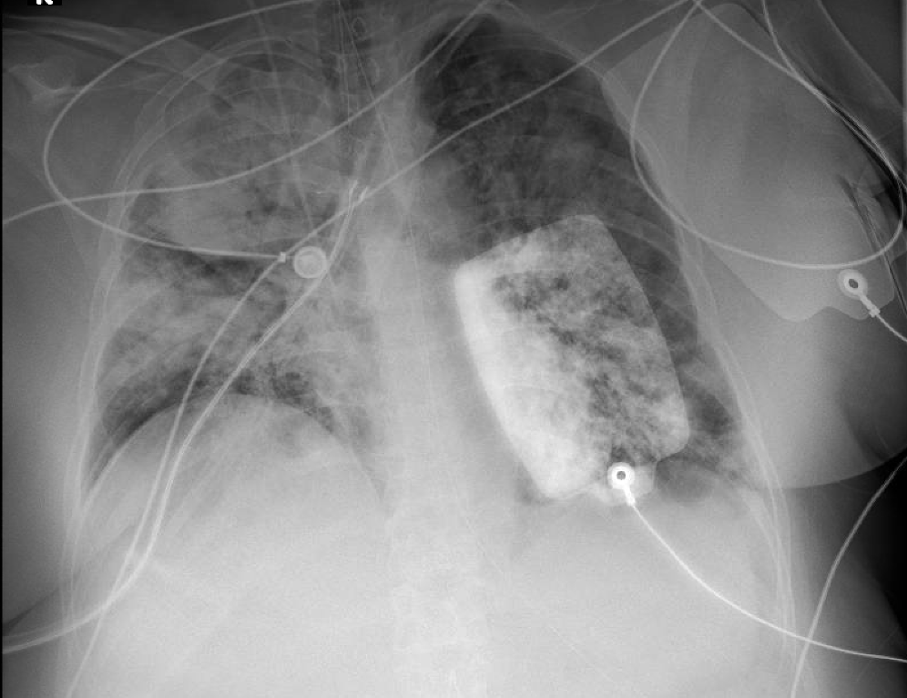
Patient Presentation
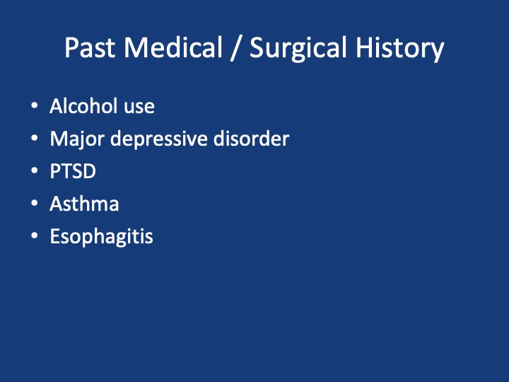
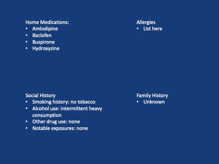
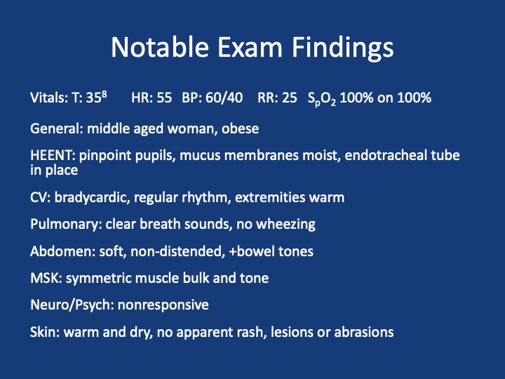
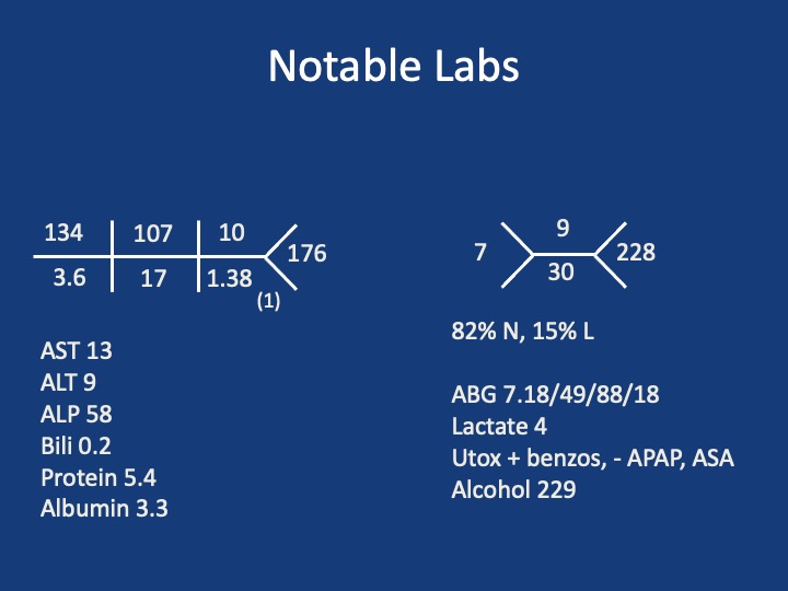
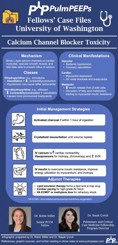
A 56-year-old woman with a history of alcohol use and depression presents after being found down at home by her boyfriend with an unknown downtime. She was found to be unresponsive and in the supine position. Her physical exam did not show any obvious trauma but the paramedics did note vomitus on her face. She received 1 L of crystalloids in the field and was intubated and brought to the ED for further management. A bag of pill bottles was found and brought with her. Her home medications include amlodipine, baclofen, buspirone, and hydroxyzine.
Key Learning Points
**Spoilers Ahead** If you want to think through the case on your own we advise listening to the episode first before looking at the infographic below
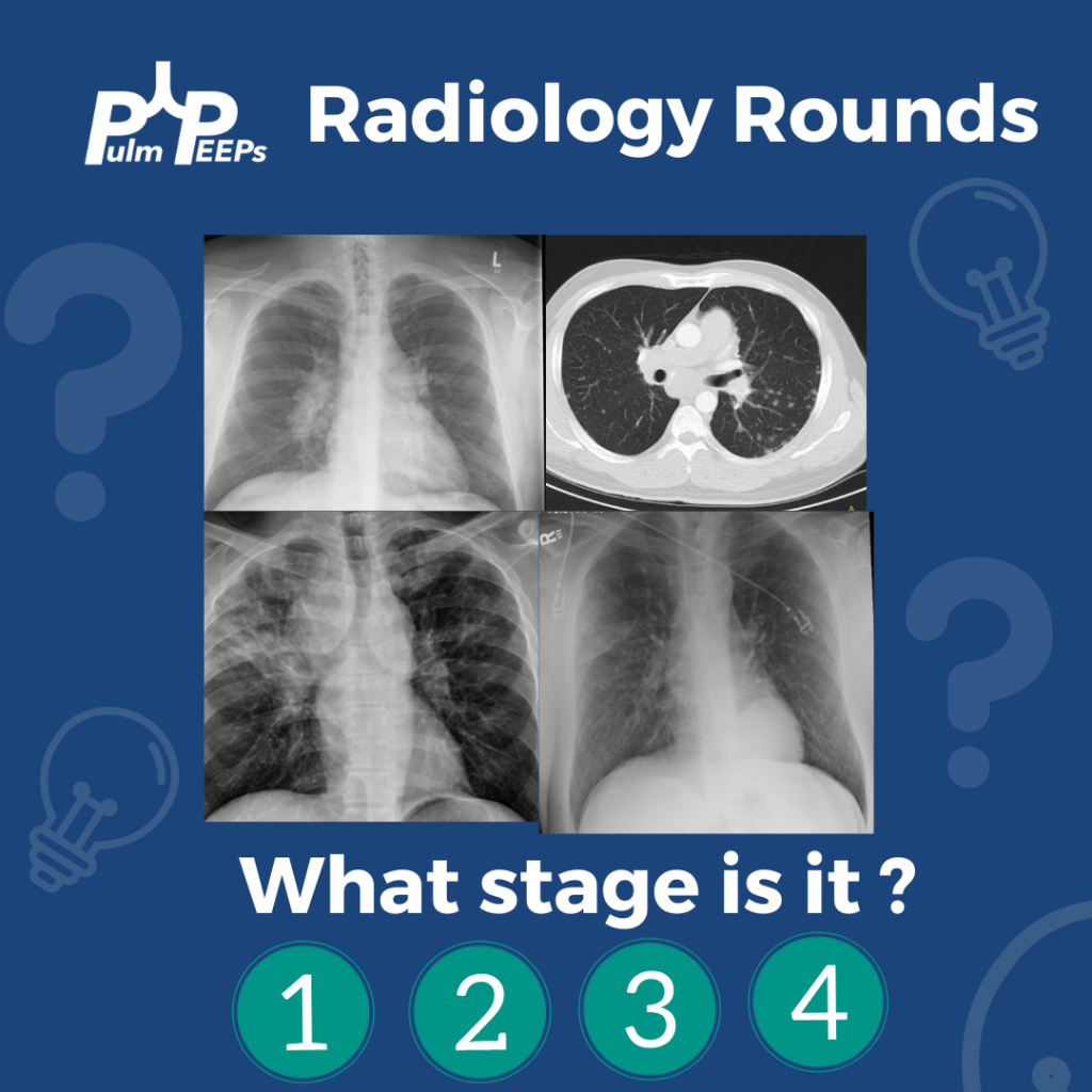
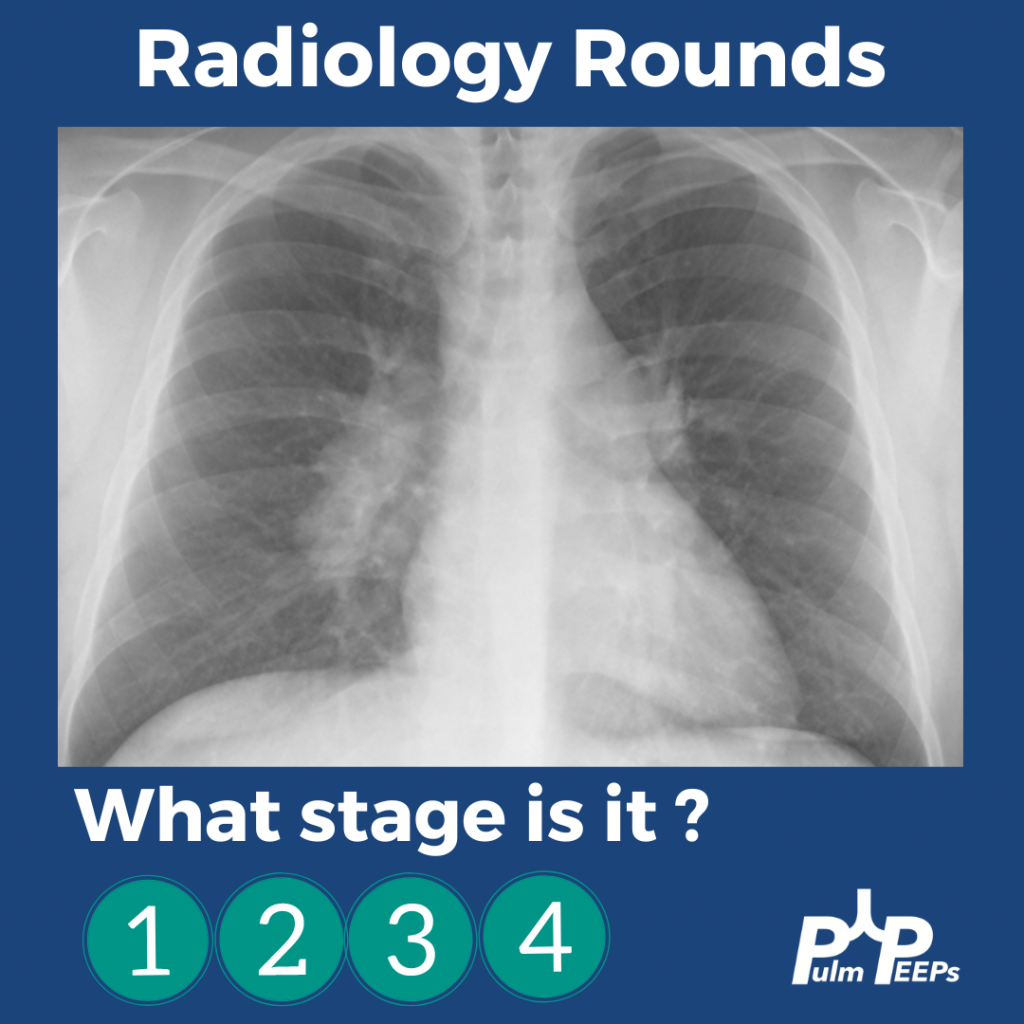
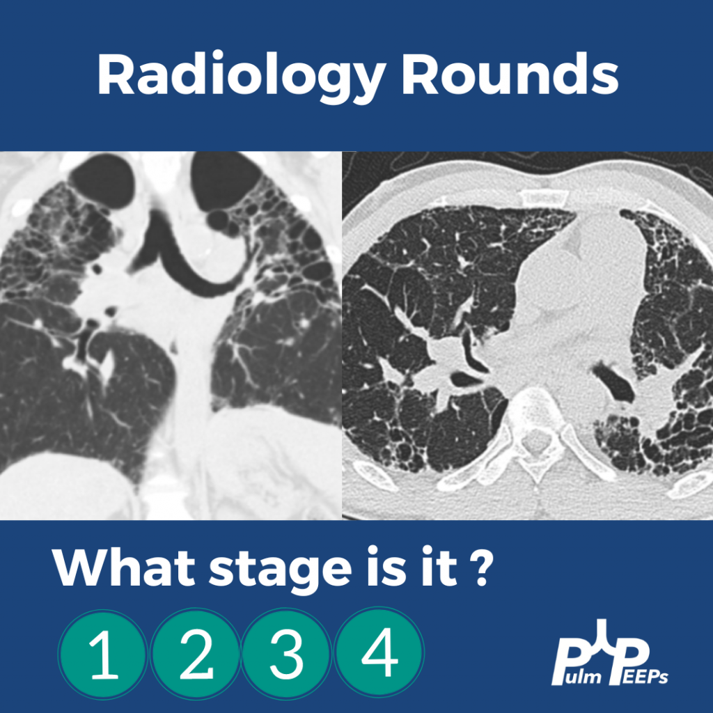
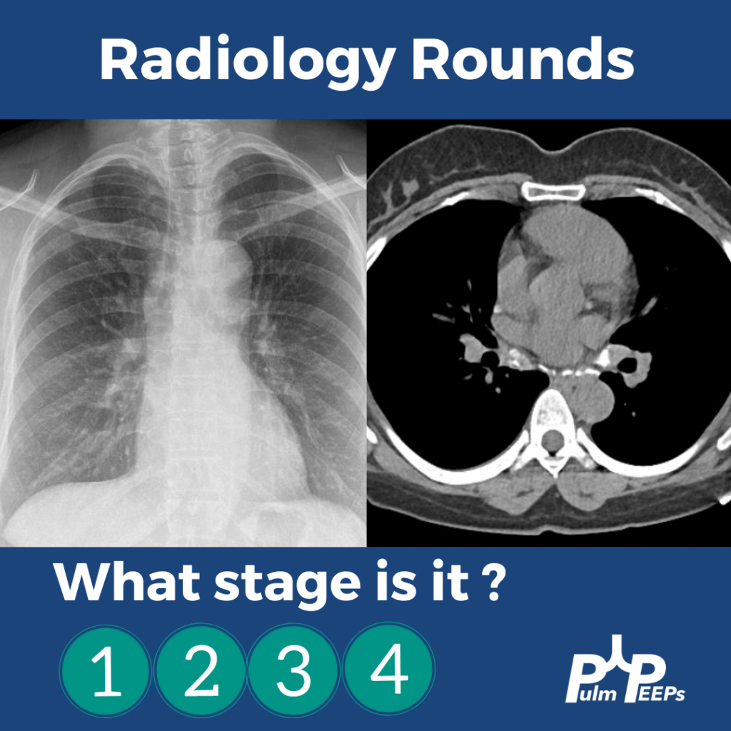
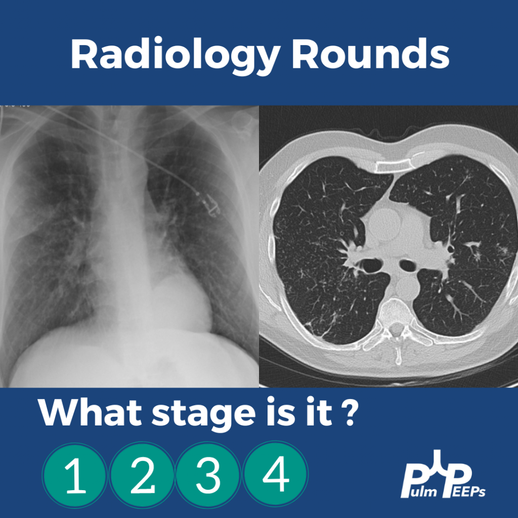
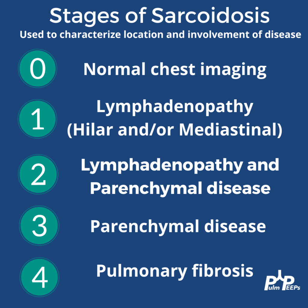
It is Tuesday and we have another Radiology Rounds we can’t wait to share with you. Follow along and see if you select the right answer as we go through different presentations of sarcoidosis and pick your answer! What stage is it?!
A middle-aged man presents to you after he was found to have hilar adenopathy on a routine chest x-ray.
A middle-age man presents with dyspnea on exertion, night sweats and weight loss. You see evidence of bilateral apical disease, and fibrosis with evidence of honeycombing on chest CT.
A young woman presents with dyspnea on exertion and was found to have hilar adenopathy with parenchymal disease.
An elderly man presents with dyspnea on exertion and was found to have nodular parenchymal disease without extensive lymphadenopathy.
This week we are absolutely thrilled to be launching a new series here at Pulm PEEPs. This is the first episode in our new Fellows’ Case Files series. The purpose of this series is to highlight the incredible clinical work that is done by pulmonary and critical care fellows everywhere, share fascinating cases from across the world, and assemble a diverse network of pulmonary and critical care educators. For each episode, we will visit a different institution, and be joined by a current fellow and the Pulmonary and Critical Care Fellowship Program Director. Our aim is to learn from them, amplify some incredible teaching points, and hear about their program. We hope you enjoy it, and if you have a case you want to bring on the series reach out to us on Twitter or at our email pulmpeeps@gmail.com.
Meet Our Guests
Fahid Alghanim is a senior pulmonary and critical care fellow at the University of Maryland. He attended medical school at the Lebanese American University Gilbert and Rose-Marie Chagoury School of Medicine and completed his internal medicine residency at Johns Hopkins Bayview. He has published on topics ranging from lung transplants to patient navigators in the ICU.
Dr. Van Holden is an Associate Professor of Medicine at the University of Maryland School of Medicine and the Pulmonary and Critical Care Fellowship Program director. Clinically, she specializes in interventional pulmonology. She is also an accomplished educator and is very active with the American Thoracic Society. She helped write the 2021 Critical Care Core Curriculum and helped coordinate the 2022 Resident Boot Camp.
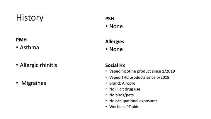
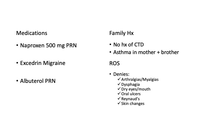
Patient Presentation
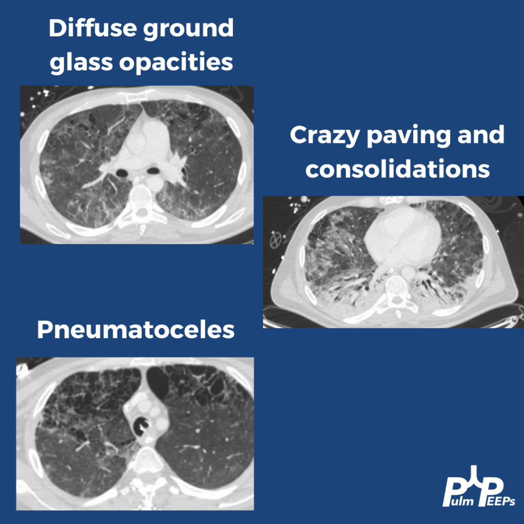
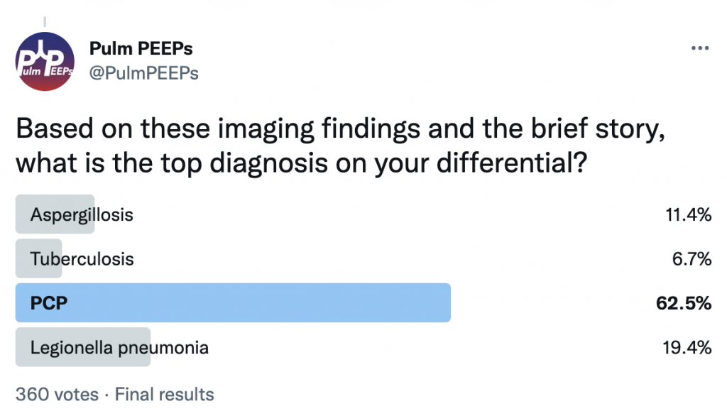
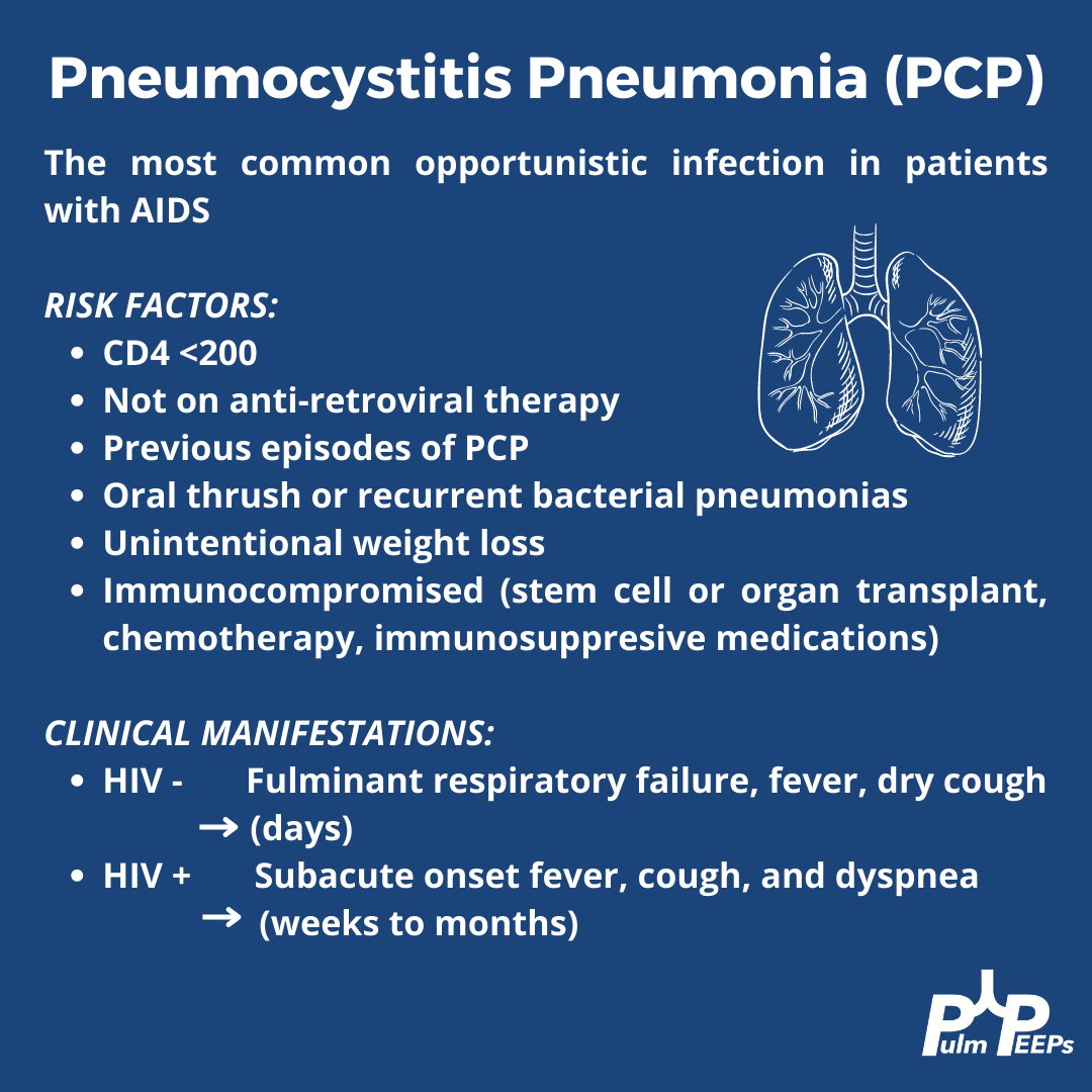
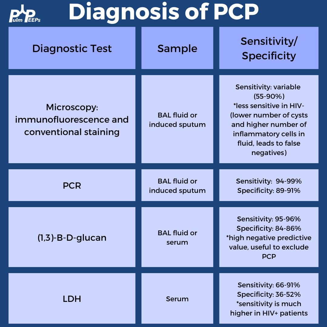
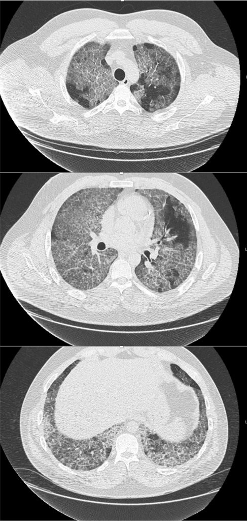
A 26-year-old man presents to his primary care doctor with 1.5 months of intermittent dyspnea, cough, chest tightness, and fatigue. His dyspnea was initially exertional, and he noticed he could do less at the gym. However, in the past 3-4 weeks it has progressed to being even with mild movement. His brother was recently diagnosed and treated for acute bronchitis so he thought this could be similar. In the office, he is noted to be tachypneic with an oxygen saturation of 83% breathing ambient air. A chest X-ray is obtained and he is sent urgently to the emergency department.
Key Learning Points
**Spoilers Ahead** If you want to think through the case on your own we advise listening to the episode first before looking at the infographics below
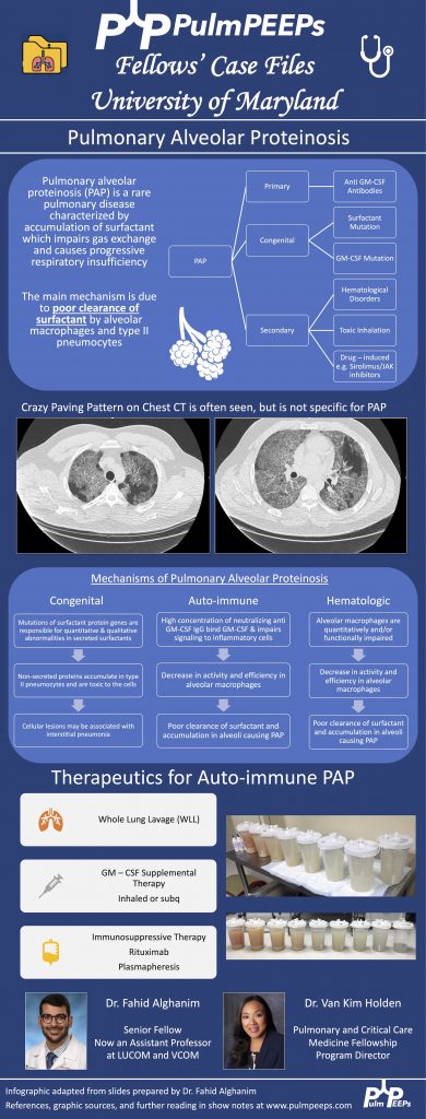
Crazy Paving is a radiological term describing ground glass opacities with superimposed interlobular septal thickening. The differential diagnosis is broad and includes infectious, neoplastic, and autoimmune processes. It is not limited to just Pulmonary alveolar proteinosis (PAP) but is suggestive in an appropriate clinical setting.
PAP is a disorder of surfactant production or clearance and its etiology is divided into three major subgroups. Primary or autoimmune; Secondary such as from toxic inhalations, hematological disorders, or medications; and Congenital
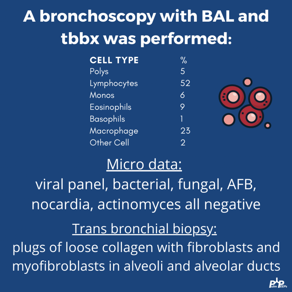
PAP is diagnosed by positive Periodic acid-Schiff (PAS) staining of lipo-proteinaceous material in the distal bronchioles and alveoli on lung biopsy. The diagnosis can be made with PAS-positive BAL staining, but this has limited sensitivity and lung biopsy is necessary for the diagnosis in up to 30 – 35% of cases.
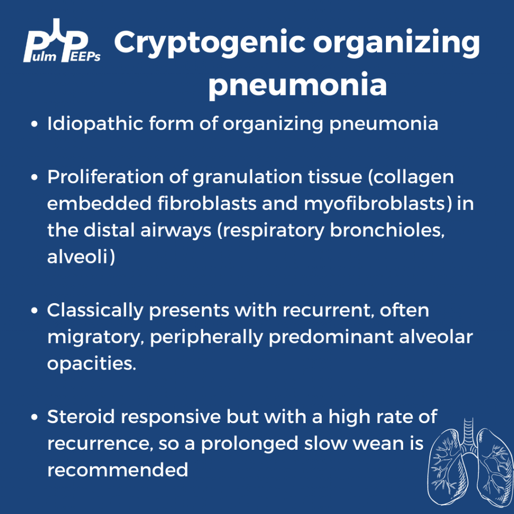
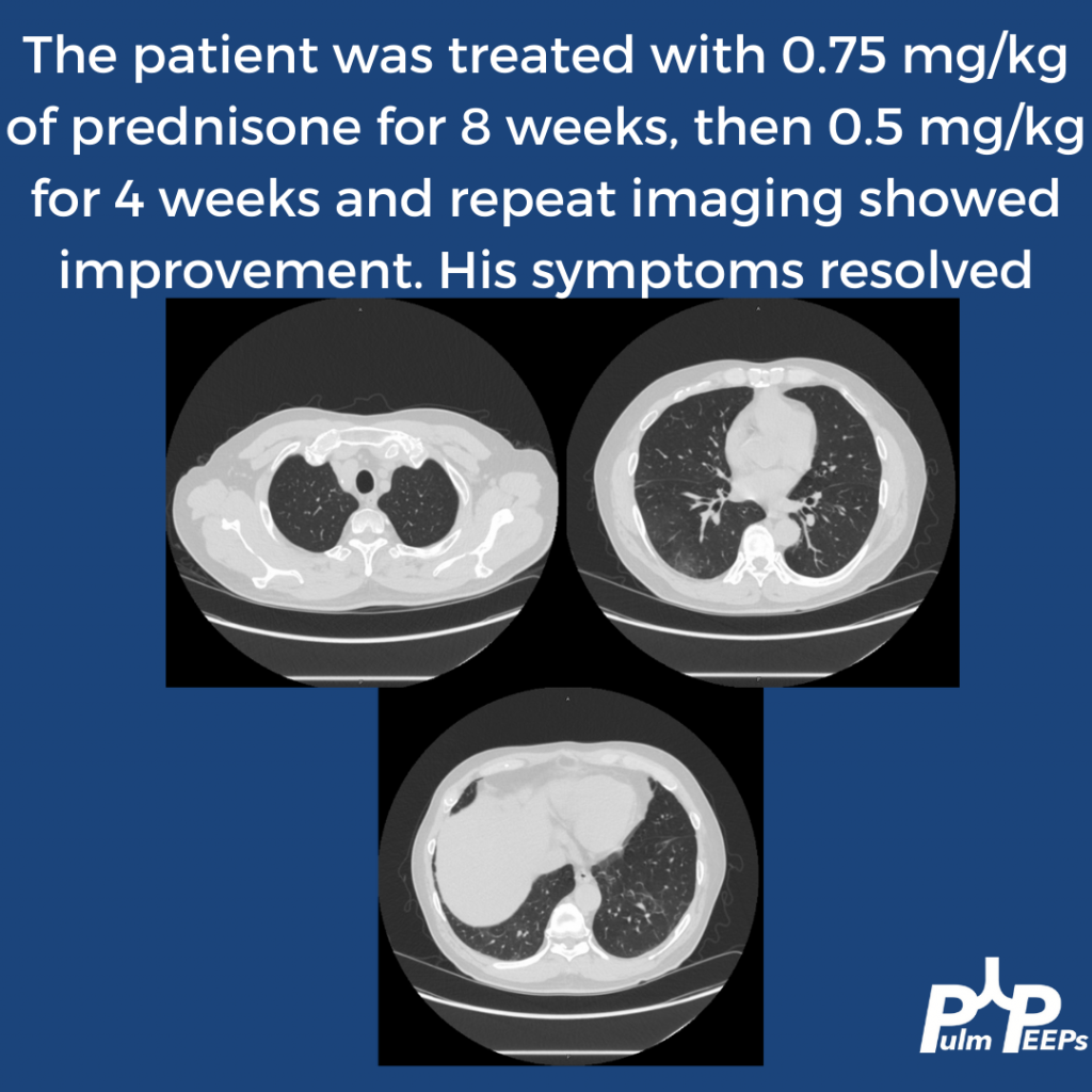
It is important not to anchor on a diagnosis when a patient presents to you for re-evaluation even if seen by a prior expert. This was pivotal in this case!
Please don’t put anything in your lung. Any toxic inhalation exposure could result in significant damage to lung parenchyma and morbidity as a result.
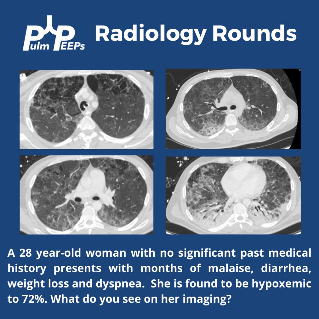
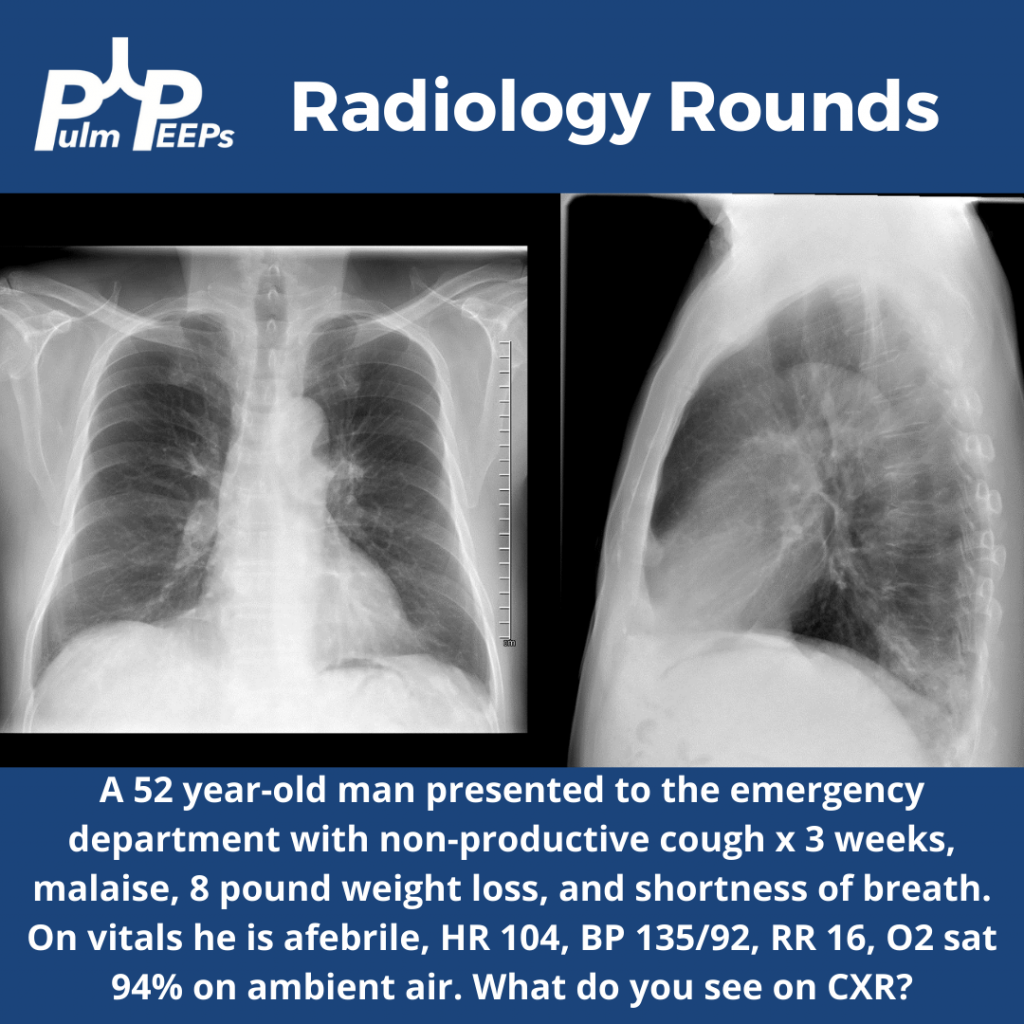
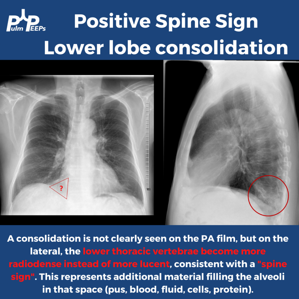
It is Tuesday and time for another #RadiologyRounds!! This is a patient who presented to the emergency department with symptoms of cough, dyspnea, malaise, and weight loss. A PA and lateral CXR was obtained.
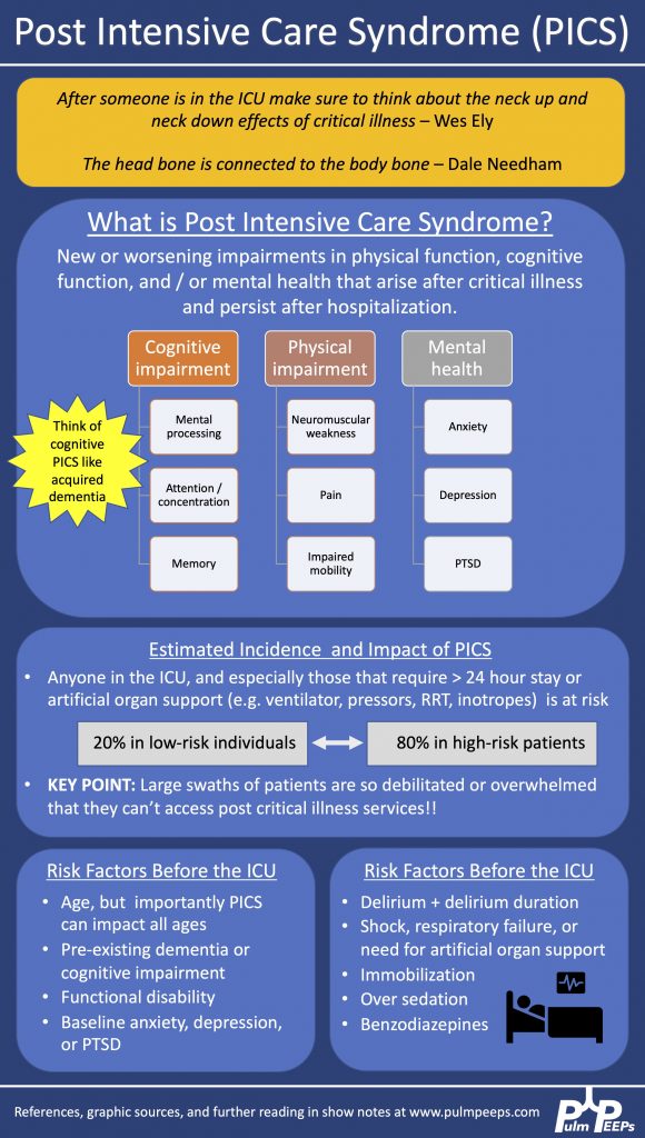
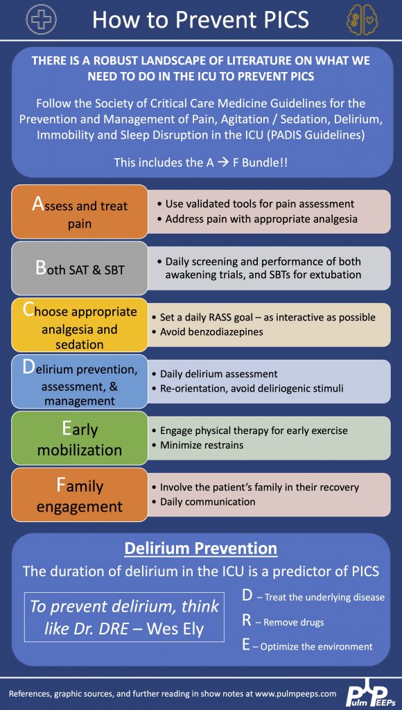
Today on Pulm PEEPs, we are joined by two pioneers in the field of post-intensive care outcomes and delirium research. Drs. Dale Needham and Wes Ely talk to us all about the Post Intensive Care Syndrome (PICS) and cover everything from how it was first recognized, to the impact it has, and, most importantly, what we can do to prevent it. This is a huge topic in the field of critical care and we’re thrilled to be delving into it with such knowledgeable guides.
Meet Our Guests
Wes Ely is the Grant W. Liddle Chair in Medicine and a Professor of Medicine at Vanderbilt University Medical Center. He is also the Associate Director of Aging Research at the VA Tennessee Valley Geriatric Research and Education Clinical Center and the co-director of the Critical, Illness, Brain Dysfunction and Survivorship Center. He has published 100s of manuscripts on critical illness survivorship and delirium. He also published a book called “Every Deep-Drawn Breath” about his and his patients’ experiences in the ICU and about the ramifications of critical illness. All net proceeds for the book are going to the CIBS Center Endowment for Survivorship
Dale Needham is a Professor of Medicine at Johns Hopkins, where he is also the Medical Director of the Critical Care Physical Medicine and Rehabilitation Program and the Director of the Outcomes After Critical Illness and Surgery Group. He is the author of 100s of publications focusing on post-ICU outcomes and has received numerous research grants from the NIH and other organizations.
Key Learning Points
Visit our website www.pulmpeeps.com to see the key learning points from this episode summarized in two infographics.
Time for another #RadiologyRounds!! This week we’re looking at the coronal CT scan views, which can be extremely helpful and are often under-utilized. Follow us on Twitter to work through Radiology Rounds cases as they come out.
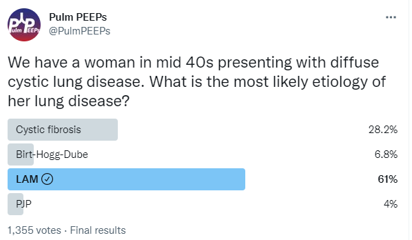
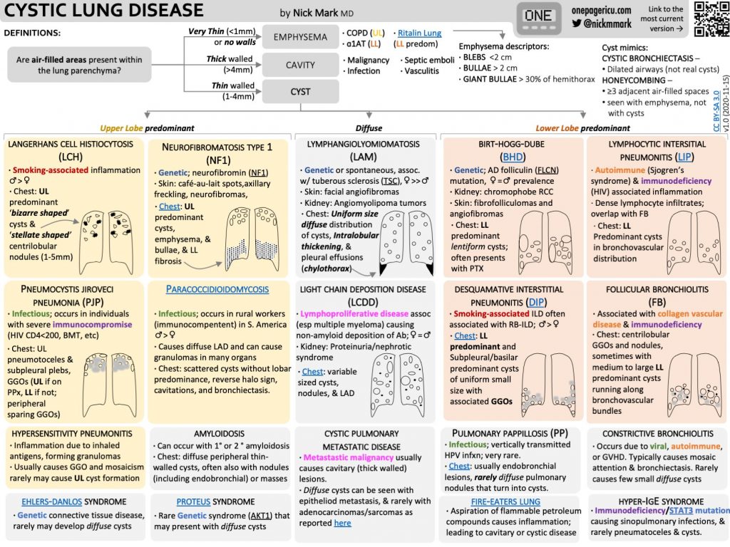
Our patient had an elevated VEGF-D level, a renal angiomyolipoma identified on CT abdomen, and imaging with diffuse cystic lung disease confirming her diagnosis of LAM. Make sure to check out the ICUOnePager made by Dr. Nick Mark.
This week on Pulm PEEPs, we are continuing our Top Consults series with a discussion on the work-up and diagnosis of Pulmonary Hypertension. See our prior Radiology Rounds on signs of PAH on CT scan, and listen to our follow-up episode on right heart catheterizations for some background before this episode… or dive right in! We’ll cover everything from history and physical, to recent guideline changes in the definition of PH, and much, much more!
Meet Our Guests
Erika Berman Rosenzweig is a Professor of Pediatrics and the Director of the Pulmonary Hypertension Center and CTEPH Program at Columbia University Medical Center / New-York Presbyterian Hospital. She is an active member of the Pulmonary Hypertension Association, was the Editor-in-Chief of Advances in Pulmonary Hypertension and is on the Scientific Board of the World Symposium on PH.
Catherine Simpson is an Assistant Professor of Medicine at Johns Hopkins Hospital and is one of the faculty members in our Pulmonary Hypertension group. Her clinical and research areas of expertise are in pulmonary vascular disease and right heart function. Her research is focused on novel biomarker discovery and metabolomics in pulmonary vascular disease.
Cyrus Kholdani is an Instructor in Medicine at Beth Israel Deaconess Medical Center and Harvard Medical School. He is also the director of the Pulmonary Hypertension Program at BIDMC, and is actively involved in clinical care and clinical research in a variety of pulmonary vascular disease domains.
Consult Patient
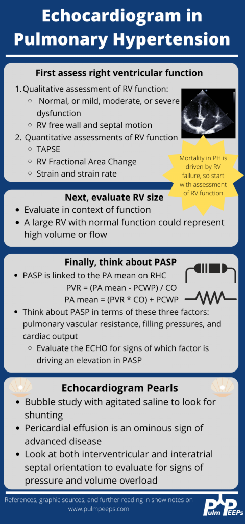
Ms. Pamela Harris (PH) is a 47-year-old woman with PMH of migraines, obesity s/p gastric sleeve (BMI now 33), and a history of remote DVT in her 20s while on OCP s/p 6 months of AC who is referred to pulmonary hypertension clinic for evaluation of dyspnea on exertion. She has actually had dyspnea for some time and previously it has been attributed to her weight. Based on this, she pursued a gastric sleeve and has lost 55 pounds, but continues to have shortness of breath. She has no cough, and does not get dyspnea at rest, but notes that after 1 flight of stairs, or 2-3 blocks on flat ground she has shortness of breath. She saw her PCP and had basic labs, basic spirometry, and an echocardiogram. He did not note anything significant on examination in the notes.
The labs had no anemia, and normal renal and liver function. Her serum bicarbonate was 25 and there was no blood gas. Spirometry showed an FVC 82% predicted, FEV1 83% predicted, and FEV1/FVC was 99% predicted. The echocardiogram had normal LVEF, mild LVH, normal RV size and function qualitatively. There was mild TR with tricuspid valve peak regurgitant velocity of 3.4 m/sec. The estimated PASP + RA pressure (based on normal IVC diameter 2.1 cm) was 46 mmHg.
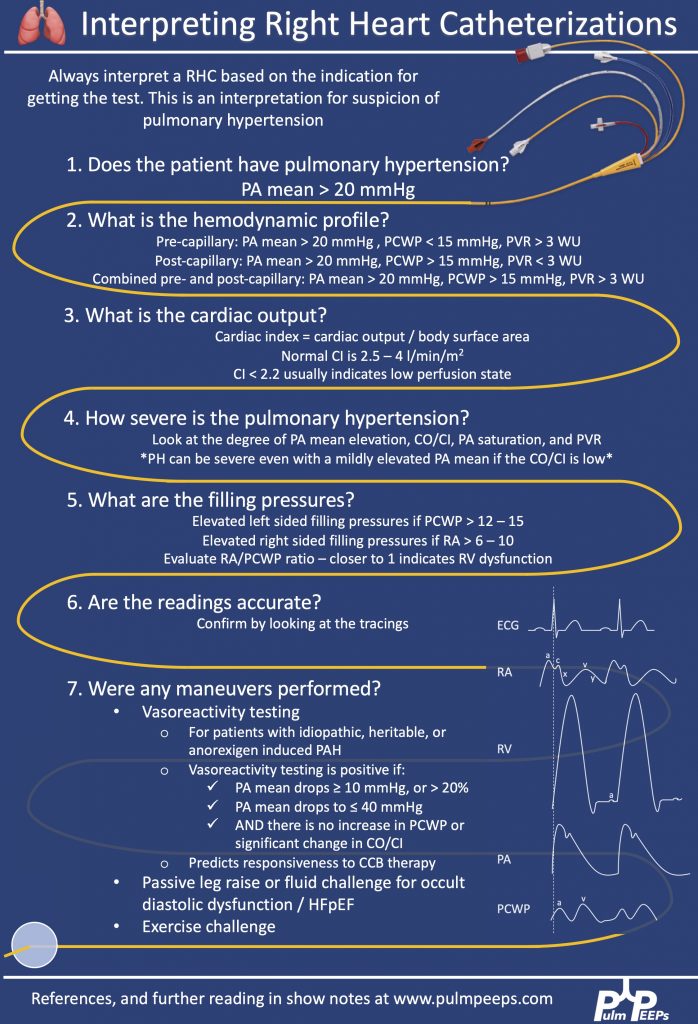
RHC: Systemic BPs 140s/90s, with O2 saturations 97-98% on RA throughout. RA mean pressure was 9, RV was 48 with an RVEDP of 17, PA was 48/27 with mean of 34, and PCWP mean was 11. CO/CI by Fick was 5.56 / 2.42, and by thermo was similar, 5.8 / 2.52. Her PA sat was 62%, and PVR was 3.97 WU.
Key Learning Points
History
Understand the constellation of symptoms and the functional limitation
The goal is to assign a WHO functional class by the end of the visit
Evaluate the time course and evolution of the symptoms
Concerning symptoms that need to be addressed
Palpitations
Pre-syncope
Syncope
Chest pain
LE edema
Evaluate for risk factors to explain or contribute to pulmonary hypertension
Signs or symptoms of OSA
Signs or symptoms of auto-immune disease
Raynauds
Skin changes
Family history
Heritable lung disease
Clotting disorders
Auto-immune disease
Social history
Exposure history
Smoking
Physical Exam
Look for signs that confirm PH
Loud P2
Accentuated with elevated PVR
Can hear pretty early on. Could be one of the earliest findings
TR murmur – pansystolic murmur at RUSB
Diastolic murmur if severe pulmonary insufficiency
Look for signs of right heart failure
JVD
S4 gallop – later in course
RV heave – later in course
Peripheral edema
Pulsatile liver or hepatosplenomegaly
Look for signs of other secondary causes of PH
Mitral regurgitation or aortic stenosis murmur
Asymmetric lower extremity edema
Pulmonary edema
Skin findings concerning for auto-immune disease or liver disease
Arthritis
Work up for etiology of PH
CBC with diff – myeloproliferative and hemolytic anemia
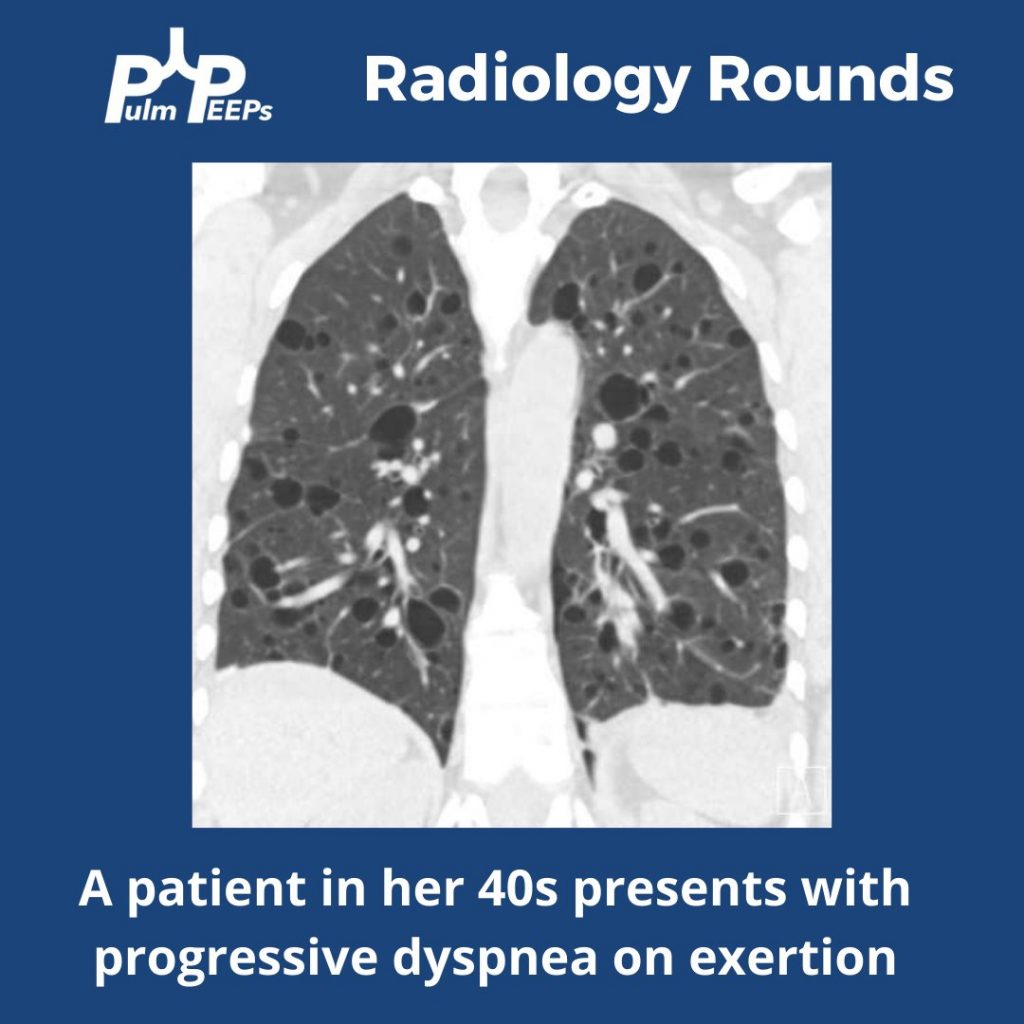
This week’s #RadiologyRounds is brought to you by our newest Contributor, Matthew Tsai! Matt will be continuing to work with us to bring you great cases and images and we are thrilled to have him on the team! Follow us on Twitter and Instagram for our Radiology Rounds, podcast episode releases, and more!